CASE PRESENTATION
Fournier's gangrene: a highly invasive necrotizing fasciitis. Case report
Gangrena de fournier: una fascitis necrotizante de alto grado de invasividad. Presentación de un caso
José Alfredo Gallego-Sánchez1* https://orcid.org/0000-0002-7686-8776
Reynaldo López-Milanés1 https://orcid.org/0000-0001-9270-9604
Yoandry Valdés-Infante2 https://orcid.org/0000-0001-5883-0697
Alejandro Román-Rodríguez3 https://orcid.org/0009-0008-6349-7161
1 University of Medical Sciences of Las Tunas. Puerto Padre Medical Sciences Branch. Puerto Padre, Las Tunas, Cuba.
2 University of Medical Sciences of Las Tunas. Guillermo Domínguez López General Teaching Hospital. Puerto Padre, Las Tunas, Cuba.
3 University of Medical Sciences of Havana. Calixto García Faculty. Havana, Cuba.
* Corresponding author: jg97@nauta.cu
Received: 22/03/2025
Accepted: 26/05/2025
How to cite this article: Gallego-Sánchez JA, López-Milanés R, Valdés-Infante Y, Román-Rodríguez A. Fournier's gangrene: a highly invasive necrotizing fasciitis. Case report. [Internet]. 2025 [cited access date]; 5:e324. Available in: https://revmedest.sld.cu/index.php/medest/article/view/324
ABSTRACT
Introduction: Fournier's gangrene is a highly invasive necrotizing fasciitis of the perineal, genital and perianal regions, which can sometimes extend to the abdominal wall as a result of a polymicrobial infection in most cases.
Objective: to present the clinical-surgical evolution of Fournier's gangrene in a 69 years old male patient.
Case presentation: the case of a white male patient, 69 years old and from a rural background, with an individual pathological history of arterial hypertension, diabetes mellitus and ischemic heart disease in the form of an acute myocardial infarction is described. He came to the consultation due to severe scrotal pain, with a change in color and foul-smelling secretions in the right hemiescrotum, which had been going on for two days. Once the diagnosis was established, taking into account the clinical data, together with the performance of complementary tests such as complete blood count and soft tissue x-ray, emergency surgery was decided, which was successful, however, the patient did not recover. of the toxic infectious condition and on the fifth day of evolution in the Intensive Care Unit, he died.
Conclusions: Fournier's gangrene is a highly invasive infection. The case of a patient who presented with clinical and paraclinical characteristics of this disease was presented, who died on his fifth postoperative day.
Keywords: Case reports; Diagnosis; Fournier's gangrene; Necrotizing fasciitis; Postoperative; Surgical procedures
RESUMEN
Introducción: la gangrena de Fournier es una fascitis necrotizante de un alto grado de invasividad de las regiones perineal, genital y perianal, que en ocasiones puede extenderse hasta la pared abdominal como resultado de una infección polimicrobiana en la mayoría de los casos.
Objetivo: presentar la evolución clínico quirúrgica de la gangrena de Fournier en un paciente masculino de 69 años de edad.
Presentación del caso: se describe el caso de un paciente masculino, de piel blanca, de 69 años de edad y de procedencia rural, con antecedentes patológicos individuales de hipertensión arterial, diabetes mellitus y cardiopatía isquémica a forma de infarto agudo del miocardio. Acudió a consulta por presentar fuertes dolores escrotales, con cambio de coloración y secreciones fétidas en el hemiescroto derecho, de dos días de evolución. Una vez establecido el diagnóstico, teniendo en cuenta los datos clínicos, unidos a la realización de exámenes complementarios como hemograma completo y radiografía de partes blandas, se decidió su intervención quirúrgica de urgencia, la que resultó exitosa, sin embargo, el paciente no se recuperó del cuadro tóxico infeccioso y al quinto día de evolución en la Unidad de Cuidados Intensivos, falleció.
Conclusiones: la gangrena de Fournier es una infección de un alto grado de invasividad. Se presentó el caso de un paciente que cursó con características clínicas y paraclínicas de esta enfermedad, el cual falleció en su quinto día de postoperatorio.
Palabras clave: Diagnóstico; Fascitis necrotizante; Gangrena de Fournier; Informes de casos; Postoperatorio; Procedimientos quirúrgicos
INTRODUCTION
Fournier's gangrene is characterized by a highly invasive necrotizing fasciitis of the perineal, genital, and perianal regions (1,2) and even the abdominal wall (2), resulting in a polymicrobial infection in most cases. (1)
Contrary to what one might think, it is not a modern nosological entity. Its first descriptions date back to 877 AD, attributed to the Persian physician Avicenna. However, it was not until 1883 that Dr. Jean Alfred Fournier gave it its current name: Fulminant gangrene of the penis and scrotum. (2)
Fournier's gangrene is a rare infection, (3) with an incidence of 1.6 cases per 100 000 men per year (4) and a mortality rate ranging from 7.8% to 50 %. (3) The average age of onset is 50 years, but it occurs in a very wide range, from 42 to 70 years, so the risk of presentation increases proportionally with age. (1)
Due to its fulminant evolution (progression of 2-3 cm/hour) (1), it constitutes a medical-surgical emergency requiring immediate intervention and subsequent management in the Intensive Care Unit.
Due to the severity of this infection, its low incidence, and the need to understand the key elements for establishing its diagnosis and emergency surgical treatment, this article aims to present the clinical and surgical evolution of Fournier's gangrene in a 69-year-old male patient.
CASE PRESENTATION
A 69-year-old Caucasian male from a rural area had a history of high blood pressure for six years, for which he had been on regular treatment with captopril (25 mg), one tablet every eight hours, and hydrochlorothiazide (25 mg), one tablet daily; and ischemic heart disease in the form of an acute myocardial infarction approximately four years prior, for which he had been on treatment with nitrosorbide (10 mg), one tablet every eight hours, and aspirin (81 mg), one tablet daily. He also suffered from type 2 diabetes mellitus and was treated with 40 units of slow-acting insulin in the morning and 20 units at night, plus 10 units of simple insulin at breakfast, lunch, and dinner. He reported that his blood glucose levels remained around 18 mmol/L. Four months ago, he suffered from acute otitis media due to herpes zoster, which caused peripheral facial paralysis.
He came to the doctor because he had been experiencing pain in his scrotum for two days, initially with pruritus. After the pruritus, the pain and volume of the right hemisphere increased, with changes in skin color. The following day, he began experiencing yellowish, foul-smelling scrotal discharge. He reported no fever, but he did experience loss of appetite and fatigue.
Physical examination:
Deviation of the corner of the mouth to the right.
Male genital system: enlargement of the right hemisphere with changes in color and loss of skin continuity, presence of foul-smelling purulent secretions; subcutaneous emphysema at the scrotum level.
Based on the data obtained from the history and physical examination, Fournier's gangrene was suspected, so urgent complementary tests were performed. The patient presented anemia, with a hematocrit of 0,36 L/L. A white blood cell count (WBC) revealed leukocytosis of 21,4 per 109/L, predominantly polymorphonuclear cells (91 %) and lymphocytes (9 %). Blood glucose was 17,6 mmol/L. Creatinine was measured, and the values were within normal limits (82 mmol/L).
A soft tissue x-ray of the scrotum showed increased radiolucency in these tissues, confirming the presence of air at this level.
The results of the laboratory and imaging studies, combined with the clinical context and history, led to a final diagnosis of Fournier's gangrene. The approach in this case was initially to stabilize the patient with hydration with 500 mL of physiological saline (0,9 %), half an ampule of polysalt (25,6 mEq), and an ampule of magnesium sulfate (10 %) for seven days. Broad-spectrum antimicrobials were prescribed for 14 days with ceftazidime (1 g) as an intravenous bulb every 8 hours, clindamycin (600 mg/4 mL) as an intravenous bulb every 12 hours, and amikacin (500 mg/2 mL) as an intravenous bulb daily for 14 days. Surgical treatment was then used.
Emergency surgical treatment was performed (Figure 1) under the following protocol:
1) Anesthetic management: Initial spinal anesthesia, followed by general orotracheal anesthesia for the main procedure.
2) Open cystostomy: Placement of a 16 Fr Foley catheter, properly secured for urinary diversion.
3) Necrectomy and scrotal debridement: Wide excision of necrotic tissue, with complete exposure of both testes and spermatic cords. Exhaustive lavage with 500 mL of saline solution (0,9 %) to remove debris and infected material.
4) Preparation of the surgical site: Repeat lavage with sterile saline solution and iodine to reduce bacterial load. Topical dressing: Application of nitrofurazone cream (0,2 %/25 g) and packing with sterile gauze.
5-) Management of septic shock: Administration of epinephrine (adrenaline) as a vasoactive amine: Dose: 3 ampoules (1 mg/mL each) in 500 mL of saline solution (0,9 %) as a continuous infusion.
6-) Strict hemodynamic monitoring for patient stabilization
Once the surgery was completed, a consultation with the Intensive Care Unit and transfer to the unit were requested.
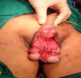
Image 1. Result of the patient's surgery
(Image by the authors).
In the days following surgery, daily dressing was performed (Figure 2) to remove the limited underlying necrotic tissue with some sloughing, and the patient responded favorably to surgical treatment. However, the infectious toxicity worsened, and symptoms of multiorgan failure manifested. Finally, on the fifth postoperative day, the patient died in the Intensive Care Unit.
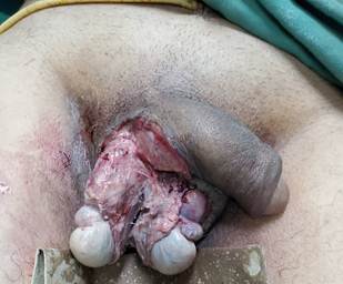
Image 2. Daily cure (Image by the authors).
DISCUSSION
Defects in the scrotal region are most often caused by Fournier's gangrene (5). This condition is a necrotizing fasciitis that produces extensive cellulitis with severe involvement of the subcutaneous tissue (6). Regarding its etiology, Flores et al. (1), Pérez et al. (3), and Iglesias et al. (6) agree on its polymicrobial nature; however, there have been cases in which only one germ was identified by culture, as was the case of Önder et al. (7), who described the first case of Fournier's gangrene associated with Rothia dentocariosa, highlighting the variability with which germs are presented as the cause of the disease.
According to Escudero et al. (4), the most frequently isolated germs are Escherichia coli in more than 50 % of cases, Bacteroides ssp. in 47 %, Streptococcus ssp. In 42 % of patients, Staphylococcus spp. in 27 %, and Enterobacter ssp. in 27 % of patients; this contradicts the results of Calderón et al. (8), who stated that Pseudomona aeruginosa was the most frequently present germ in cultures. This case coincided with the previous authors' findings, as the isolated germs were Escherichia coli and Streptococcus ssp.
According to Flores et al. (1), the age groups in which Fournier's disease most frequently occurs range from 42 to 70 years, and although both sexes can be affected, it is more common in males. This data was consistent with those of this article, as the patient was 69 years old and male.
Flores et al. (1) consider that there are several predisposing factors for the development of Fournier's gangrene, each of which has an underlying cause: a decrease in the body's immune capacity. In this case, the patient is a hypertensive patient who also suffers from type 2 diabetes mellitus, whose blood glucose levels usually remain above normal, creating a favorable environment for the reproduction of various microorganisms.
Regarding the presentation of this disease, Sandoval et al. (2) and Iglesias et al. (6) concluded that it depends on the stage of the infection, the patient's comorbidities, and their general health. Initially, there may be a prodromal period, during which the patient may experience general malaise, itching, and fever for several days or even weeks, after which more severe symptoms begin. These symptoms include increased scrotal or vulvar pain, which generally does not correspond to clinical findings, edema, cellulitis, and erythema, which may be accompanied by a foul odor, crepitus, and systemic symptoms such as fever, low blood pressure, and tachycardia.
These data correspond to those obtained from the patient's physical examination and questioning, and the patient waited three days to seek medical attention. This delay in seeking medical attention, in the authors' opinion, allows for the proliferation of cell colonies that invade the tissues, which favors the infectious toxicity and hinders the patient's prognosis.
Although the diagnosis is primarily clinical, Iglesias et al. (6) consider that laboratory, imaging, and histopathological examinations help confirm the diagnosis. This is why Escudero et al. (4) recommend taking a complete medical history, including a detailed list of medical history. These authors recognize the importance of imaging studies to complement the diagnosis. Plain abdominal or pelvic radiographs are relevant as they allow the presence of gas in the abdominal wall to be observed, making surgery absolutely necessary.
Other highly relevant imaging studies include ultrasound, which delineates soft tissue involvement at the genital level, avoiding delays in the diagnosis of this disease and differentiating it from other surgical emergencies such as testicular torsion. Computed tomography, according to Escudero et al. (4), is currently considered the primary imaging tool, especially when deep tissue or retroperitoneal space involvement is suspected. Pérez et al. (3) state that gadolinium-enhanced magnetic resonance imaging is another excellent method for examining soft tissue.
In this case, the diagnosis was established based on the results of the physical examination and history, as well as plain radiography, which coincides with these criteria. The authors consider that, although a correct anamnesis guides the diagnosis towards Fournier's gangrene, urgent imaging and analytical studies should be performed to confirm the diagnosis.
Regarding treatment, Pérez et al. (3) include several pillars such as parenteral hydration of the patient, administration of broad-spectrum antimicrobials intravenously, surgical debridement of all necrotic tissue, treatment of the infectious focus and, in some cases, hemodynamic stabilization of the patient. Escudero et al. (4) include hydroelectrolytic and nutritional management within the treatment, in addition, they establish a mixed parenteral antibiotic therapy regimen, recommending the administration of three medications such as first or third generation cephalosporins, metronidazole or clindamycin and an aminoglycoside. Once the patient is stabilized, maintaining the same with broad-spectrum triple antibiotic coverage, surgical debridement of non-viable necrotic tissue should be performed, in a broad manner, until reaching areas of healthy tissue, removing any area suspected of infection (3,4).
The therapeutic approach to the patient described in this article was consistent with that proposed by these authors, yielding good results. Once the wound base is clean and free of signs of infection, and covered by granulation tissue, reconstruction can be considered. Bravo and González (9) suggest mobilizing a full-thickness flap, a myocutaneous flap, a cutaneous flap, or tackling the wound edges.
Regarding survival, Flores et al. (1) report that in 75 % of cases, patients with Fournier's gangrene die. This figure may increase with the increase in the infectious toxicity, even when surgery is successful in more than 75 % of cases. This was consistent with the present article, where, despite the patient's successful surgery, the infectious toxicity worsened, and the patient died.
CONCLUSIONS
Fournier's gangrene is a highly invasive infection. We present the case of a patient with clinical and paraclinical features of this disease. Surgery combined with broad-spectrum antimicrobial therapy is the treatment of choice for this condition. Following surgery, the patient progressed satisfactorily; however, the toxic infection was unable to be reduced, resulting in death.
BIBLIOGRAPHIC REFERENCES
1. Flores-Galván KP, Aceves-Quintero CA, Guzmán-Valdivia G. Gangrena de Fournier. Rev Cir Gen [Internet]. 2021 [cited 21/03/2025]; 43(2):107-114. Available in: http://www.scielo.org.mx/scielo.php?script=sci_arttext&pid=S1405-00992021000200107&Ing=es. https://doi.org/10.35366/106721
2. Sandoval J, Aldana C, Balmelli B. Carácterísticas epidemiológicas y quirúrgicas en pacientes con secuelas de enfermedad de Fournier. An Fac Cienc Méd. (Asunción) [Internet]. 2023 [cited 21/03/2025]; 56(3):67-75. Available in: http://scielo.iics.una.py/scielo.php?script=sci_arttext&pid=S1816-89492023000300067&Ing=en. https://doi.org/10.18004/anales/2023.056.03.67
3. Pérez-Ladrón de Guevara P, Cornelio-Rodríguez G, Quiroz-Castro O. Gangrena de Fournier. Reporte de caso. Rev Fac Med Méx [Internet]. 2020 [cited 21/03/2025]; 63(5): 26-30. Available in: http://www.scielo.org.mx/scielo.php?script=sci_arttext&pid=S0026-17422020000500026&Ing=es. https://doi.org/10.22201/2448486e.2020.63.5.04
4. Escudero-Sepúlveda AF, Cala-Durán JC, Belén-Jurado M, Tomasone SE, Carlino Currenti VM, Abularach Borda R et al. Conceptos para la identificación y abordaje de la gangrena de Fournier. Rev Colomb Cir [Internet] 2022 [cited 21/03/2025]; 37(4):653-664. Available in: http://www.scielo.org.co/scielo.php?script=sci:arttext&pid=S2011-75822022000400653&Ing=en. https://doi.org/10.30944/20117582.930
5. Ramírez-Antúnez P, Aldana C, Peña A, Berra P. Reconstrucción escrotal con colgajo pediculado del músculo gracilis bilateral e injerto de piel parcial. An. Fac. Cien. Méd. (Asunción) [Internet]. 2023 [cited 21/03/2025]; 56(1):103-8. Available in: http://scielo.iics.una.py/scielo.php?script=sci_arttext&pid=S1816-89492023000100103&Ing=en. https://doi.org/10.18004/anales/2023.056.01.103
6. Iglesias-Guzmán MH, del Carpio JM, Bustamante-Lozada A, de Pawlikowski-Amiel NW, Caller-Farfán V, Medina-Castillo JM. Experiencia y manejo de dos casos de gangrena de Fournier en pacientes pediátricos en el Instituto Nacional de Salud del Niño-San Borja. Acta méd. Perú [Internet]. 2021 [cited 21/03/2025]; 38(4):319-323. Available in: http://www.scielo.org.pe/scielo.php?script=sci_arttext&pid=S1728-59172021000400319&Ing=es. http://dx.doi.org/10.35663/amp.2021.384.2134
7. Önder T, Alkan S, Tezcan S. A case of Fournier's gangrene caused by Rothia dentocariosa. Iberoam J Med [Internet]. 2023 [cited 21/03/2025]; 5(2): 84-7. Available in: http://scielo.isciii.es/Scielo.php?script=sci_arttext&pid=S2695-50752023000200005&Ing=es. https://dx.doi.org/10.53986/ibjm.2023.0012
8. Calderón W, Camacho J, Obaíd M, Moraga J, Bravo D, Calderón D. Tratamiento quirúrgico de la gangrena de Fournier. Rev Cir [Internet]. 2021 [cited 21/03/2025]; 73(2): 150-157. Available in: http://www.scielo.cl/scielo.cl/scielo.php?script=sci_arttext&pid=S2452-45492021000200150&Ing=es. http://dx.doi.org/10.35687/s2452-45492021002748
9. Bravo-Gálvez VM, González-Villegas HO. Reconstrucción de las secuelas de la gangrena de Fournier, reporte de dos casos. Rev Mex Urol [Internet]. 2020 [cited 21/03/2025]; 80(6): e09. Available in: http://www.scielo.org.mx/scielo.php?script=sci_arttext&pid=S2007-40852020000600009&Ing=es. https://doi.org/10.48139/revistamexicanadeurologa.v80i6.648
AUTHORSHIP STATEMENT
JAGS: conceptualization, formal analysis, investigation, methodology, visualization, writing - original draft, writing - review & editing.
RLM: conceptualization, formal analysis, investigation, methodology, writing - original draft, writing - review & editing.
YVI: conceptualization, formal analysis, investigation, methodology, visualization, writing - original draft, writing - review & editing.
ARR: conceptualization, formal analysis, investigation, methodology, visualization, writing - original draft, writing - review & editing.
CONFLICTS OF INTEREST
The authors declare no conflicts of interest.
FUNDING SOURCES
The authors did not receive funding for this research.