CASE PRESENTATION
Extraction of the upper central incisor and spontaneous eruption of an impacted canine: Case report
Exodoncia del incisivo central superior y erupción espontánea de canino retenido: Reporte de caso
Mónica González Guerra 1*, https://orcid.org/0000-0001-9861-3267
Ana Flavia Cundú Zayas 1, https://orcid.org/0009-0009-3374-8510
Damarys Calvo Pérez 2, https://orcid.org/0000-0002-6275-4647
1 Teaching Stomatological Clinic 3rd PCC Congress. Matanzas, Cuba
2 Eliseo Noel Caamaño Pediatric Hospital. Matanzas, Cuba
* Corresponding author: monicagonzalezguerra96@gmail.com
Received: 24/07/2025
Accepted: 25/12/2025
How to cite this article: González-Guerra M; Cundú-Zayas AF; Calvo-Pérez D. Extraction of the upper central incisor and spontaneous eruption of an impacted canine: Case report. MedEst. [Internet]. 2025 [cited access date]; 5:e467. Available in: https://revmedest.sld.cu/index.php/medest/article/view/467
Abstract
Introduction: Impacted maxillary canines are among the most frequent eruptive disorders in orthodontics, often leading to root resorption of adjacent incisors and functional or esthetic complications.
Objective: To describe a clinical case of an impacted right maxillary canine associated with severe root resorption of the central incisor, its surgical management, and therapeutic decision.
Case report: A patient presented with mobility of the right maxillary central incisor (11). Radiographic examination revealed severe root resorption of 11 caused by the ectopic eruption path of the right maxillary canine (13). Extraction of 11 was performed, and spontaneous eruption of 13 was allowed. Clinical and radiographic follow-up showed favorable eruptive progress.
Conclusions: Early diagnosis of impacted canines is crucial to prevent severe resorption of adjacent teeth. Therapeutic decisions must be individualized, balancing surgical intervention and the potential for spontaneous eruption.
Keywords: Impacted Canine; Root Resorption; Orthodontics; Case Report
RESUMEN
Introducción: Los caninos maxilares retenidos constituyen una de las alteraciones eruptivas más relevantes en ortodoncia, con potencial de provocar reabsorción radicular de los incisivos adyacentes y complicaciones funcionales y estéticas.
Objetivo: Describir un caso clínico de retención del canino superior derecho con reabsorción radicular del incisivo central, su abordaje quirúrgico y la decisión terapéutica adoptada.
Presentación del caso: Paciente que acude por movilidad dentaria del incisivo central superior derecho (11). La radiografía evidenció pérdida de continuidad radicular del 11 asociada a la posición ectópica del canino superior derecho (13). Se realizó exodoncia del 11 y se decidió dejar al 13 erupcionar espontáneamente. El seguimiento clínico-radiográfico mostró una adecuada respuesta erupcional.
Conclusiones: El diagnóstico precoz de los caninos retenidos es esencial para prevenir reabsorciones radiculares graves. La decisión terapéutica debe individualizarse considerando la posición del diente, el daño a estructuras vecinas y la posibilidad de erupción espontánea.
Palabras clave: Canino Retenido; Reabsorción Radicular; Ortodoncia; Caso Clínico
INTRODUCTION
Impacted teeth represent an eruption disturbance that can lead to local complications, (1,2) including root resorption and functional and aesthetic alterations. Among these, maxillary canines are the third teeth most frequently impacted, after third molars and second premolars. (3,4)
Several studies have demonstrated the importance of early diagnosis, (5,6) using imaging techniques such as CBCT, which allow for assessment of the canine's position and the degree of involvement of adjacent teeth. (7,8)
The therapeutic management of impacted canines includes alternatives such as surgical exposure with orthodontic traction, extraction, and, in selected cases, waiting for spontaneous eruption. (9,10) The choice depends on the patient's age, the degree of associated resorption, functional viability, and the aesthetics of the affected tooth. (3,11)
The decision to treat an impacted canine by waiting for its eruption (observation) versus a surgical approach depends on several factors: the canine must have a favorable inclination with minimal deviation from its normal eruptive path, there must be sufficient space in the dental arch or it can be created with orthodontics, there must be no associated pathologies (such as cysts, ankylosis, or resorption of adjacent teeth), and the patient must be young enough to have a high potential for spontaneous eruption (7).
Advantages of Observation (Waiting for Eruption):
• Less invasive.
• Lower cost.
• Tissue preservation.
• Less stress for the patient.
• Possible spontaneous eruption.
A well-documented clinical case on waiting for the eruption of an impacted canine is essential for several reasons.
1. Education and Learning: It allows for sharing experience and developing skills.
2. Research and Evidence: Ensures the generation of important data and the identification of patterns.
3. Improvement of Clinical Practice: Provides the establishment of practical guidelines and a scientific-clinical basis for the evaluation of treatments.
4. Interdisciplinary Communication: Establishes a common language that facilitates effective communication among different specialists.
This paper aims to present a clinical case of retention of the upper right canine associated with severe root resorption of the central incisor, in which a conservative therapeutic approach was chosen to promote spontaneous eruption.
CASE PRESENTATION
A 13-year-old male patient with no significant medical history presented to the Orthodontics Clinic at the "Eliseo Noel Camaño" Pediatric Hospital in Matanzas. The reason for consultation was a dentoalveolar trauma sustained after biting his brother, according to his mother. The incident occurred 24 hours prior to the evaluation. This episode resulted in concussion of the affected dental tissues, dental luxation, and extreme tooth mobility.
Clinical findings:
Intraoral examination revealed permanent dentition in the upper arch, with pathological mobility of the right maxillary central incisor (tooth 11). The clinical absence of the right maxillary permanent canine (tooth 13) and insufficient space in the arch for its proper eruption were noted.
Radiographic findings:
A periapical radiograph of the upper anterior region was requested, which revealed root resorption of tooth 11 (Figure 1). This resorption was attributed to the presence of the impacted canine (13) dental follicle in an ectopic position, displacing and affecting the incisor root.
Therapeutic management and outcome:
The patient was referred to the Maxillofacial Surgery Service with the initial plan to perform surgical exposure of the canine (13), place an orthodontic appliance (bracket), and apply guided orthodontic traction. However, during the surgical procedure (Figure 2), after the extraction of the crown of incisor 11, which had a guarded prognosis, the clinical team reconsidered their strategy (Figure 3). Given the patient's young age and the final position of the canine after the extraction of tooth 11, a wait-and-see approach was chosen to encourage its spontaneous eruption.
Follow-up appointments were performed to monitor the case's progress, initially every 3 months and subsequently at 6 months.
After the first 3 months, a periapical radiograph was taken to assess the canine's eruptive movement and complete root integrity.
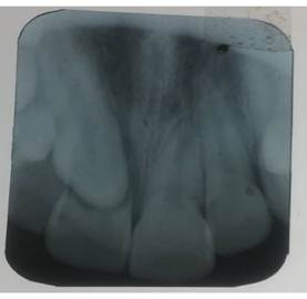
Figure 1. Periapical radiograph of the upper anterior sector.
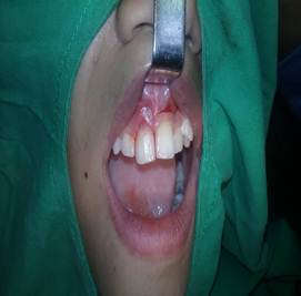
Figure 2. Surgical procedure
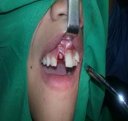
Figure 3. Extraction of tooth 11.
DISCUSSION
Impacted maxillary canines are a common finding in orthodontic practice, with a reported prevalence between 1% and 3%, predominantly affecting females and the maxillary arch. (1, 3) While this condition is common, root resorption of a maxillary central incisor as a complication, as seen in this case, is a less frequent event, but of great clinical relevance due to its functional and aesthetic implications. (4, 6)
Previous studies agree that the proximity of the impacted canine to the incisor roots significantly increases the risk of root resorption, as demonstrated by the study by Li N et al. (4). Ericson S et al. (12) quantified this risk, showing that up to 12% of lateral incisors and 2% of central incisors may exhibit resorption associated with this condition. (12)
For accurate diagnosis, cone-beam computed tomography (CBCT) has become the preferred tool, as it allows for the early identification of resorption areas and precise evaluation of the canine's three-dimensional position. (13,14) While authors such as Güllü YÖ et al. (1) have indicated that position and angulation on panoramic radiographs are useful predictors of therapeutic success, recent literature supports CBCT as the optimal diagnostic standard in these cases. (6,7)
In the patient presented, the periapical radiograph confirmed the loss of root continuity of the central incisor (11), a finding that proved crucial in treatment planning.
After extraction of tooth 11, the canine's condition was reassessed (13). Given its favorable inclination, acceptable eruption trajectory, absence of associated pathology, sufficient space in the arch—a condition favored by the previous extraction—and a high potential for spontaneous eruption due to the patient's age, a watchful waiting approach was chosen, allowing for natural eruption.
Treatment may include surgical exposure with orthodontic traction, extraction of the impacted tooth, or allowing spontaneous eruption. (9,10) In this case, spontaneous eruption was chosen, a course of action supported by reports such as that of Selmani et al. (3), who describe successful cases under favorable conditions of space and tooth position. Likewise, recent reviews recommend individualizing the treatment plan, considering the associated risks. (11,12)
The watchful waiting approach adopted in this case is supported by the literature, which recognizes spontaneous eruption as a viable option when the canine has a favorable trajectory and there is adequate space in the dental arch. (8) Becker A et al. (15) support this approach, reporting success rates exceeding 70% after surgical exposure and subsequent guided eruption.
The resolution of these complex cases invariably requires an interdisciplinary approach, as emphasized by Mousa MR et al. (14). Collaboration between orthodontists and maxillofacial surgeons is crucial to optimize aesthetic and functional outcomes. In this context, the extraction of incisors with severe resorption—far from representing a therapeutic failure—should be considered a necessary measure to preserve the integrity of the supporting tissues. (16)
This clinical case coincides with the findings of Becker A et al. (17), who demonstrated that early extraction of severely affected teeth, followed by surgical exposure of the impacted canine, can offer a favorable prognosis with rigorous follow-up. The experience of the authors of this study reinforces the validity of this protocol.
The therapeutic approach adopted in this clinical case is consistent with current evidence. The preservation of the permanent canine and the long-term function of the anterior sector were prioritized through an individualized decision based on clinical and radiographic findings.
CONCLUSIONS
Retained maxillary canines can cause severe complications in adjacent teeth, such as root resorption. Early diagnosis and the use of advanced imaging techniques are essential for treatment planning. This case demonstrates that, under favorable conditions, spontaneous eruption of the retained canine is a valid alternative that should be considered in orthodontic practice.
BIBLIOGRAPHIC REFERENCES
1. Güllü YÖ, Çakmak Özlü F. Prediction of the success of orthodontic treatment of impacted maxillary canines using panoramic radiography parameters: retrospective cross-sectional study. BMC Oral Health. [Internet] 2024 [cited 10/07/2025]; 24:1547. Available in: https://pubmed.ncbi.nlm.nih.gov/39719561/
2. Halim H, Halim IA. Management of Unilateral Impacted Maxillary Permanent Canine: A Case Report. Open Dent J. [Internet] 2023 [cited 10/07/2025]; 18:e18742106298043. Available in: https://www.opendentistryjournal.com/VOLUME/18/ELOCATOR/e18742106298043/FULLTEXT/
3. Selmani M, Duci B, Ismaili S. Orthodontic and Surgical Management of Impacted Maxillary Canines: A Narrative Review. Eur J Gen Dent. [Internet] 2024 [cited 10/07/2025]; 13(3):177-182. Available in: https://papers.ssrn.com/sol3/papers.cfm?abstract_id=5499778
4. Li N, Yang L, Yang Q, Wang H, Xu X, Wang T. Long-term follow-up after the treatment of impacted canines in the maxilla causing severe root resorption of the lateral incisors: two case reports. BMC Oral Health. [Internet] 2024 [cited 10/07/2025]; 24(1):482. Available in: https://pubmed.ncbi.nlm.nih.gov/38643143/
5. Ng WL, Cunningham A, Pandis N, Bister D, Seehra J. Impacted maxillary canine: Assessment of prevalence, severity and location of root resorption on maxillary incisors: A retrospective CBCT study. Int Orthod. [Internet] 2024 [cited 10/07/2025]; 22(3):100890. Available in: https://pubmed.ncbi.nlm.nih.gov/38838434/
6. Alshawy ES, Kolarkodi SH. Revealing the Effect of Impacted Canines on the Adjacent Teeth. A Three Dimensional Study Using CBCT. J Pharm Bioallied Sci. [Internet] 2023 [cited 10/07/2025]; 15(Suppl 1):S720-S724. Available in: https://pubmed.ncbi.nlm.nih.gov/37654346/
7. Hirschhaut M, Leon N, Gross H, Flores-Mir C. Guidance for the Clinical Management of Impacted Maxillary Canines. Compend Contin Educ Dent. [Internet] 2021 [cited 10/07/2025]; 42(5):220-228. Available in: https://pubmed.ncbi.nlm.nih.gov/33980019/
8. Hirschhaut M, Weinstein C, Flores Mir C, Naoumova J. Orthodontic management of ectopic and impacted teeth. Semin Orthod. [Internet] 2025 [cited 10/07/2025]; 31(1):34-45. Available in: https://www.sciencedirect.com/science/article/pii/S1073874625000349
9. Ceraulo S, Barbarisi A, Oliva B, et al. Treatment Options in Impacted Maxillary Canines: A Literature Review. Dent J (Basel). [Internet] 2025 [cited 10/07/2025]; 13(9):433. Available in: https://pubmed.ncbi.nlm.nih.gov/41002706/
10. Lwin CT, Cooney M, Goh M, Tham D, Nowak S. Factors Associated With Successful Surgical Exposure of Impacted Maxillary Canines. J Oral Maxillofac Surg. [Internet] 2024 [cited 10/07/2025]; 82(1):93-101. Available in: https://pubmed.ncbi.nlm.nih.gov/37683693/
11. Balasuppramaniem MT, Anitha A, Manovijay B, Ravi S. Various surgical methods of impacted maxillary canine exposure: A case series. J Indian Soc Periodontol. [Internet] 2023 [cited 10/07/2025]; 27(2):212-215. Available in: https://pubmed.ncbi.nlm.nih.gov/37152453/
12. Ericson S, Kurol J. Resorption of incisors after ectopic eruption of maxillary canines: a CT study. Angle Orthod. [Internet] 2000 [cited 10/07/2025]; 70(6):415-23. Available in: https://pubmed.ncbi.nlm.nih.gov/11138644/
13. Walker L, Enciso R, Mah J. Three-dimensional localization of maxillary canines with cone-beam computed tomography. Am J Orthod Dentofacial Orthop. [Internet] 2005 [cited 10/07/2025]; 128(4):418-23. Available in: https://pubmed.ncbi.nlm.nih.gov/16214621/
14. Mousa MR, Hajeer MY, Burhan AS, Heshmeh O. Adult periodontal comparison after treatment of palatally impacted canines aligned by conventional or accelerated minimally-invasive corticotomy-assisted orthodontic traction: A randomized controlled trial. Int Orthod. [Internet] 2023 [cited 10/07/2025]; 21(3):100785. Available in: https://pubmed.ncbi.nlm.nih.gov/37329591/
15. Becker A, Chaushu S. Success rate and duration of orthodontic treatment for adult patients with palatally impacted maxillary canines. Am J Orthod Dentofacial Orthop. [Internet] 2003 [cited 10/07/2025]; 124(5):509-14. Available in: https://pubmed.ncbi.nlm.nih.gov/14614417/
16. Oberoi S, Knueppel S. Three-dimensional assessment of impacted canines and root resorption using cone beam computed tomography. Oral Surg Oral Med Oral Pathol Oral Radiol. [Internet] 2012 [cited 10/07/2025]; 113(2):260-7. Available in: https://pubmed.ncbi.nlm.nih.gov/22677744/
17. Becker A, Chaushu S. Surgical Treatment of Impacted Canines: What the Orthodontist Would Like the Surgeon to Know. Oral Maxillofac Surg Clin North Am. [Internet] 2015 [cited 10/07/2025]; 27(3):449-458. Available in: https://pubmed.ncbi.nlm.nih.gov/26231817/
AUTHORSHIP STATEMENT
MGG: Conceptualization, Data Curation, Formal Analysis, Research, Methodology, Resources, Supervision, Validation, Visualization, Writing, Writing - Review and Editing.
AFCZ: Conceptualization, Research, Resources, Visualization, Writing - Review and Editing.
DCP: Conceptualization, Formal Analysis, Research, Methodology, Resources, Supervision, Visualization, Writing - Review and Editing.
CONFLICT OF INTEREST
The authors declare no conflicts of interest.
FUNDING SOURCES
The authors received no funding for the development of this article.