CASE PRESENTATIONS
Manson's schistosomiasis in adults: presentation of five cases and literature review
Piosis de Mansoni en adultos: presentación de cinco casos y revisión de la literatura
Alvaro Rojas Mora 1*, https://orcid.org/0000-0001-6805-5364
Graciela Fundora Guerra 1, https://orcid.org/0009-0000-9273-0403
Aylet Pérez López 1, https://orcid.org/0000-0003-4629-9995
1 General Calixto García University Hospital. Havana, Cuba.
* Corresponding author: alvarorojasmorapiel@gmail.com
Received: 18/07/2026
Accepted: 23/09/2026
Published: 25/09/2026
How to cite this article: Rojas-Mora A; Fundora-Guerra G; Pérez-López A. Manson's schistosomiasis in adults: presentation of five cases and literature review. MedEst. [Internet]. 2026 [cited access date]; 6:e578 Available in: https://revmedest.sld.cu/index.php/medest/article/view/578
ABSTRACT
Introduction: Mansoni’s piosis is an uncommon variant of bullous impetigo characterized by its localization in skin folds, particularly the axillae and groin. Literature on the condition is scarce and limited to isolated case reports.
Objective: To describe the clinical and epidemiological characteristics of a series of 5 cases of Mansoni’s piosis treated at the Dermatology Department of the General Calixto García University Hospital.
Case presentation: Five adult patients (3 men and 2 women), aged 26 to 56 years, with no relevant medical history, are presented. All sought consultation for bullous lesions in the axillae—present for 3 to 7 days—associated with excessive sweating and exposure to high temperatures. Physical examination revealed flaccid bullae containing purulent fluid, as well as erosions with a collarette of scales and crusts. Microbiological cultures were positive in all cases, identifying Staphylococcus aureus in 5/5 and Streptococcus pyogenes in 3/5. Combined treatment with Bidens pilosa (romerillo) compresses, topical gentamicin, and oral cephalexin proved effective, resulting in complete resolution within 8 to 12 days without complications or recurrences.
Conclusions: This series of 5 cases confirms the clinical characteristics described in classical literature but provides new observations regarding associated symptoms (pruritus and burning sensation) and mixed etiology. Microbiological confirmation in all cases and the favorable response to treatment enhance the understanding of this rare entity.
Keywords: Mansoni’s piosis; bullous impetigo; Staphylococcus aureus; Streptococcus pyogenes; case series.
RESUMEN
Introducción: La Piosis de Mansoni es una variedad infrecuente de impétigo ampolloso, caracterizada por su localización en pliegues, especialmente axilas e ingles. Su literatura es escasa y se limita a reportes de casos aislados.
Objetivo: Describir las características clínicas y epidemiológicas de una serie de 5 casos de Piosis de Mansoni atendidos en el servicio de Dermatología del Hospital Universitario General Calixto García.
Presentación del caso: Se presentan 5 pacientes adultos (3 hombres y 2 mujeres), con edades entre 26 y 56 años, sin antecedentes patológicos relevantes. Todos consultaron por lesiones ampollosas en axilas, de 3 a 7 días de evolución, asociadas a sudoración excesiva y exposición a altas temperaturas. El examen físico mostró ampollas flácidas de contenido purulento, erosiones con collarete de escamas y costras. El cultivo microbiológico fue positivo en todos los casos, identificándose Staphylococcus aureus en 5/5 y Streptococcus pyogenes en 3/5. El tratamiento combinado con fomentos de romerillo, gentamicina tópica y cefalexina oral fue efectivo, con resolución completa en 8 a 12 días, sin complicaciones ni recurrencias.
Conclusiones: Esta serie de 5 casos confirma las características clínicas descritas en la literatura clásica, pero aporta nuevas observaciones sobre la sintomatología asociada (prurito y ardor) y la etiología mixta. La confirmación microbiológica en todos los casos y la buena respuesta al tratamiento refuerzan el conocimiento sobre esta entidad infrecuente.
Palabras clave: Piosis de Mansoni; impétigo ampolloso; Staphylococcus aureus; Streptococcus pyogenes; serie de casos.
INTRODUCTION
Contagious impetigo is a superficial bacterial skin infection, highly contagious, caused primarily by Staphylococcus aureus and Streptococcus pyogenes. (1) It presents in two main clinical forms: non-bullous (or crusted) impetigo and bullous impetigo. (2)
Although it can affect people of any age, it occurs most frequently in children under 5 years of age. (1,3) Its prevalence is particularly high in tropical and subtropical regions, with rates that vary significantly depending on the setting. (3,4) Recent studies confirm that impetigo remains a global health problem, with a childhood prevalence of 12,3 % and more than 162 million children affected worldwide, with a particularly high burden in vulnerable and low-resource communities, where prevalence can reach 19,4 % in children. (4,5)
Manson's piosis is defined as an infrequent variety of bullous impetigo, characterized by its typical location in the folds, with the axillae and groin being the areas of choice. (1-3,6) It manifests with flaccid bullae, whose liquid content is initially clear and later becomes purulent. When they rupture, they leave an erosion surrounded by a collarette of scales. (6) It usually does not leave scars and may present with or without pruritus. Its course is variable: it may resolve spontaneously or require topical or systemic treatment, depending on the extent and clinical response. (1,3,6)
This entity is endemic in tropical regions and is associated with warm and humid climates, with a tendency to present in epidemic outbreaks. (5,6) It has been proposed that its pathogenesis differs from common bullous impetigo, as it is related to obstruction of the sweat duct and consequent sweat retention. (7) In this sense, excessive sweating and ductal obstruction are recognized as important risk factors. (6,7)
Despite having been documented initially in the 20th century by authors such as Simons (6) and Buist and collaborators (5), modern literature on this entity is very scarce, with only isolated case reports. (2) Recently, in 2025, a case was published in the Journal of the Colombian Association of Dermatology (2), which evidences that clinical interest in this entity still exists and that its diagnosis remains a challenge for dermatologists.
The present series of 5 cases, unlike previous isolated case reports, allows describing the clinical spectrum of Manson's piosis in a larger group of patients, identifying presentation patterns, and offering a more complete view of its course and response to treatment. (8-10)
Furthermore, microbiological confirmation is provided in all cases, which strengthens the validity of the findings and differentiates this study from previous reports based solely on clinical diagnosis. Recent reviews on the treatment of skin infections caused by Staphylococcus aureus confirm that beta-lactam antibiotics such as cephalexin remain effective and are a first-line option for the management of bullous impetigo. (8-10)
This publication aspires to illuminate a little-explored corner of dermatology, and offers clinicians a valuable tool for the early recognition of Manson's piosis. By highlighting its differential diagnosis in bullous lesions of the folds, the aim is not only to enrich the scientific body of knowledge, but also to provide tangible teaching that can be directly transferred to clinical practice, thereby improving the care of patients presenting this infrequent variety of impetigo. (2,10,12)
For these reasons, the objective of this publication is to describe the clinical and epidemiological characteristics of a series of 5 cases of Manson's piosis.
CASE PRESENTATION
A series of 5 cases of Manson's piosis is presented, treated in the Dermatology service of the General Calixto García University Hospital between January and December 2025. All patients gave their written informed consent for the publication of their clinical data and images, with preservation of their anonymity. The study was approved by the institution's Ethics Committee.
Case 1
A 42-year-old female patient, with no known personal pathological history or drug allergies. She attended the consultation due to bullous lesions in the left axilla of 5 days' evolution. She reported that the lesions appeared insidiously and spread progressively during the following days. The patient reported excessive sweating in the days prior to the appearance of the lesions, associated with household activities and exposure to high ambient temperatures.
Dermatological physical examination: Multiple bullae with purulent content were observed, on an erythematous base and with scant crusts located in the left axilla (Figure 1).
Case 2
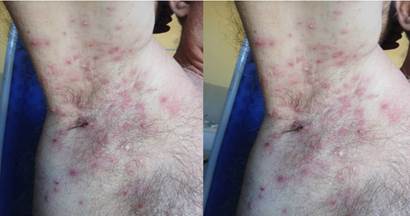
A 28-year-old male patient, with no known personal pathological history or drug allergies. He attended the consultation due to bullous lesions in the right axilla of 4 days' evolution. He reported having performed intense and prolonged physical activity in the days prior, with profuse sweating. The lesions began as small bullae that increased in number and size, and some had ruptured spontaneously. He reported mild pruritus in the affected area.
Dermatological physical examination: Multiple erosions surrounded by a collarette of scale and scant bullae with purulent content were observed, located in both axillae (Figure 2).
Case 3
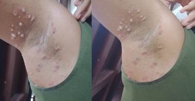
A 26-year-old male patient, with a history of previous episodes of similar bullous lesions in the same location, which resolved spontaneously, with no known drug allergies. He attended the consultation due to bullous lesions in the right axilla of 3 days' evolution, with characteristics similar to the previous episodes, but of greater extent. He reported prolonged exposure to heat and humidity in his work environment, associated with excessive sweating. He reported mild pruritus in the affected area.
Dermatological physical examination: Multiple voluminous bullae with purulent content were observed, associated with minimal crusts and erosions, located in the right axilla (Figure 3).
Case 4
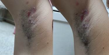
A 26-year-old female patient, with no relevant pathological history or known drug allergies. She consulted due to bullous lesions in the left axilla of 5 days' evolution. She reported that the lesions appeared suddenly and spread rapidly, and associated their appearance with excessive sweating due to household activities and exposure to high ambient temperatures.
Dermatological physical examination: Erosions surrounded by a collarette of scale and isolated purulent bullae were evidenced, located in the left axilla (Figure 4).
Case 5
A 56-year-old male patient, with no pathological history and no known drug allergies. He attended the consultation due to bullous lesions in the right axilla of 7 days' evolution. He reported that the lesions had worsened progressively, and associated their appearance with having worked in conditions of high temperature and humidity, with profuse sweating. He denied contact with people with similar lesions and had not received prior treatment. He reported mild itching and burning in the affected area.
Dermatological physical examination: Multiple bullae with purulent content and an erythematous base were observed in the right axilla, extending beyond the limits of the axillary region, with discrete crusts (Figure 5).
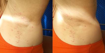
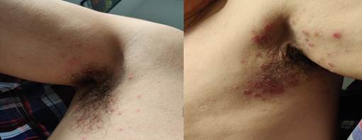
Figure 2. Multiple erosions surrounded by a collarette of scale and a few purulent blisters, located in both axillae.
Supplementary tests
All patients underwent bacteriological exudate testing of the purulent content of the bullae, processed in the microbiology laboratory of the General Calixto García University Hospital. In 100% of cases (5/5), the microbiological culture was positive, identifying Staphylococcus aureus in all cases and Streptococcus pyogenes (group A beta-hemolytic streptococcus) in 3 of the 5 cases. The antimicrobial sensitivity study showed that the isolated strains were sensitive to cephalexin and gentamicin in all cases.
Treatment and course
The treatment used in all cases consisted of a combined regimen integrating topical and systemic measures. Fresh romerillo compresses (Bidens pilosa) were used, applied twice a day on the lesions after gentle cleansing, with the aim of taking advantage of its antibacterial, anti-inflammatory, and healing properties; afterward, a thin layer of 0.1% gentamicin cream was applied over the affected areas, at a frequency of two applications daily. Additionally, as systemic treatment, all patients received cephalexin in 500 mg capsules orally, every 6 hours, for 7 days.
As part of the complementary measures, it was recommended to maintain adequate hygiene of the affected area, dry carefully to avoid maceration, avoid excessive sweating, wear loose cotton clothing, and isolate patients from direct contact with other people during the active period of the lesions. This therapeutic strategy was effective in all cases, achieving complete resolution of the lesions in a period of 8 to 12 days, without complications or recurrences during follow-up.
DISCUSSION
Manson's piosis is an infrequent variety of bullous impetigo, characterized by its typical location in the folds, especially in the axillae, as initially described by Simons. (6) The present series of 5 cases provides evidence on the clinical and epidemiological characteristics of this entity, and allows comparison of the findings with the scarce available literature, both classical and modern.
Epidemiological characteristics
Unlike common impetigo, which mainly affects children under 5 years of age, as noted by Zanoni et al. (1), Bansal et al. (3), and Wiegele et al. (4), Manson's piosis in this series presented in young and middle-aged adults, with ages ranging from 26 to 56 years. Similarly to what was described by Rodríguez-Estupiñán et al. (2) in their case report, the male sex was the most frequent in this series.
Clinical characteristics
As described by Simons in his classic study (6), the axilla was the constant location of the lesions in all cases, confirming that the folds, especially the axillae, are the characteristic site of this entity. Likewise, in the case reported by Rodríguez-Estupiñán et al. (2), the lesions were also limited to the axillary region, without involvement of the groin or other intertriginous areas.
The observed lesions—flaccid bullae with purulent content and a "collarette" border—fully coincide with Simons' description (6), who defined them as "pea-sized bullae, with turbid or purulent content, sometimes surrounded by an erythematous area." Similarly, the presence of erosions in 4 of the 5 cases and crusts in 3 of them adjusts to the natural evolution described by this author, who noted that, after rupture of the bulla, "an erosion surrounded by a collarette of scales remains." (6)
Regarding symptomatology, the absence of systemic symptoms in all cases coincides with what was noted by Simons (6), who emphasized that "there is no itching or general symptoms." However, in the present series, 3 patients reported pruritus and one of them also burning, which differs from the classic description. In contrast, the case reported by Rodríguez-Estupiñán and collaborators (2) is described as asymptomatic, which comes closer to Simons' original description. (6) This difference could be due to individual variations in the perception of pruritus or differences in the bacterial strain involved.
Risk factors
All cases reported excessive sweating in the days prior to the appearance of the lesions, associated with exposure to high temperatures and humidity. This finding is consistent with the pathophysiological hypothesis proposed by Simons (6) and supported by the studies of Sulzberger and Herrmann (7) on sweat duct obstruction, which reinforces the theory that sweat retention plays a fundamental role in the pathogenesis of the disease. This association becomes more relevant if one considers that impetigo is an infection with high prevalence in tropical and subtropical regions (3-5).
Likewise, Rodríguez-Estupiñán and collaborators (2) identified "excessive sweating in said region despite the use of deodorant" as an associated factor in their patient, which underscores the relevance of this risk factor. Furthermore, the higher prevalence of impetigo during the summer months, documented by Bansal and collaborators (3) and Wiegele and collaborators (4,5), supports the association of this entity with warm and humid climates, a finding that was also observed in the present series.
Treatment and course
The treatment applied with romerillo compresses, topical gentamicin, and oral cephalexin was effective in all cases, with complete resolution within 8 to 12 days. This result resembles what was noted by Simons (6), who described an evolution of "two or three weeks without treatment," which suggests that antibiotic therapy accelerates recovery. Similarly, Rodríguez-Estupiñán and collaborators reported resolution in their patient with a similar regimen of oral cephalexin and topical fusidic acid. (2)
Cephalexin is among the oral antibiotics recommended for bullous impetigo, particularly in extensive cases or with multiple lesions. (10,12) The sensitivity to cephalexin and gentamicin observed in all cultures supports the choice of the therapeutic regimen used. Recent reviews confirm that beta-lactam antibiotics, such as cephalexin, remain effective and constitute a first-line option. (10,12) This approach adjusts to current recommendations, which underscore the importance of systemic treatment in extensive or recalcitrant infections. (8-10,12)
As in the classic reports by Simons (6) and Barth et al., (5) no complications or recurrences were observed during follow-up in our series, which confirms the good prognosis of this entity.
Contributions of the series
The present series differs from previously published case reports (2,5,6) in that it allows a more exhaustive characterization of the disease: by covering a larger number of patients, it enables the identification of clinical patterns and offers a more comprehensive view of the course and response to treatment. Furthermore, microbiological confirmation in all cases and the detailed description of the clinical course strengthen the validity of these findings. In contrast to the report by Rodríguez-Estupiñán and collaborators (2), which is based on clinical and dermatoscopic diagnosis without microbiological confirmation, this series has microbiological cultures that confirm the etiology.
This series of 5 cases of Manson's piosis confirms the clinical characteristics described in the classical literature by authors such as Simons (6), although it provides new observations on the associated symptomatology and the mixed etiology (staphylococcus and streptococcus). Similarly to what was previously reported by Rodríguez-Estupiñán and collaborators (2), the axillary location, excessive sweating as a risk factor, and the good response to antibiotic treatment are findings consistent with our series.
On the other hand, the presence of pruritus in 3 of the 5 cases and burning in 1 of them suggests that the entity may present local symptoms that had not been previously described by Simons (6) or by other classical authors such as Buist and collaborators (5) and Castellani and Chalmers. (8)
The present series has limitations inherent to its observational design and the small sample size. Molecular typing tests of the bacterial strains were not performed, which prevents establishing phylogenetic relationships. Nor was long-term follow-up conducted to evaluate possible late recurrences. However, microbiological confirmation and the detailed description of the cases constitute strengths that contribute to the knowledge of this entity.
CONCLUSIONS
The present series of 5 cases of Manson's piosis confirms the clinical characteristics described in the classical literature, with constant axillary location and good response to combined treatment with oral cephalexin and topical gentamicin. New observations are provided on the presence of pruritus and burning in some patients, as well as the mixed etiology (Staphylococcus aureus and Streptococcus pyogenes) in most cases. Microbiological confirmation in all cases and complete resolution in 8 to 12 days without complications reinforce the good prognosis of this entity. It is recommended to consider Manson's piosis in the differential diagnosis of bullous lesions in folds of adults with risk factors such as excessive sweating and exposure to warm and humid climates.
BIBLIOGRAPHIC REFERENCES
1. Barth DD, Daw J, Xu R, Enkel S, Pickering J, McRae T, et al. Modes of transmission and attack rates of group A Streptococcal infection: a protocol for a systematic review and meta-analysis. Syst Rev. 2021;10(1):90. doi: 10.1186/s13643-021-01641-5.
2. Rodríguez-Estupiñán AC, Peña-Chaparro DA, Fonseca-López FF, Martínez-Jiménez MA. Piosis de Mansoni: correlación clínica y dermatoscópica de un caso. Rev Asoc Colomb Dermatol Cir Dermatol. 2025;33(4). doi: 10.29176/2590843X.2041.
3. Bansal R, Gupta A, Sharma P, Kumar V, Singh S. Prevalence and determinants of impetigo in rural and urban communities: a cross-sectional study. Indian J Dermatol. 2025;70(2):112-8. doi: 10.4103/ijd.ijd_124_24.
4. Wiegele S, McKinnon E, van Schaijik B, Enkel S, Noonan K, Bowen AC, et al. The epidemiology of superficial Streptococcal A (impetigo and pharyngitis) infections in Australia: a systematic review. PLoS One. 2023;18(11):e0288016. doi:10.1371/journal.pone.0288016.
5. Barth DD, Mullane MJ, Sampson C, Chou C, Pickering J, Nicol MP, Davies MR, Carapetis J, Bowen AC. Missing Piece Study protocol: prospective surveillance to determine the epidemiology of group A streptococcal pharyngitis and impetigo in remote Western Australia. BMJ Open. 2022;12(4):e057296. Published 2022 Apr 6. doi:10.1136/bmjopen-2021-057296
6. Simons RDG. Studies on Pyosis Mansoni or Impetigo bullosa tropica. Dermatologica. 1946;93(4):189-94. doi: 10.1159/000255856.
7. Sulzberger MB. The clinical significance of certain disturbances in sweating. Dermatologica. 1956 Mar;112(3):161-88. doi: 10.1159/000256313. PMID: 13330405.
8. Giacobbe DR, Dettori S, Corcione S, Vena A, Sepulcri C, Maraolo AE, et al. Emerging treatment options for acute bacterial skin and skin structure infections and bloodstream infections caused by Staphylococcus aureus: a comprehensive review of the evidence. Infect Drug Resist. 2022;15:2137-57. doi: 10.2147/IDR.S318322.
9. Thomas A, Almsallaty N, Chalati T, Boateng J, Buanz A, Totea AM. Role of methicillin-resistant Staphylococcus aureus in cutaneous infections: current treatments and therapeutic approaches for future advancement. Int J Pharm. 2025;684:126030. doi: 10.1016/j.ijpharm.2025.126030.
10. Trang DT, Buck EC, Schoenherr DT. Impetigo: rapid evidence review. Am Fam Physician. [Internet] 2026 [cited 10/07/2026];113(2). Available in: https://pubmed.ncbi.nlm.nih.gov/41839109/
11. Brazel M, Desai A, Are A, Motaparthi K. Staphylococcal Scalded Skin Syndrome and Bullous Impetigo. Medicina (Kaunas). 2021;57(11):1157. Published 2021 Oct 24. doi:10.3390/medicina57111157
12. Amgarth-Duff I, Thomas H, Ricciardo BM, Anderson L, Stephens M, Currie BJ, et al. Systematic review of the evidence for treatment and management of common skin conditions in resource-limited settings: An update. Trop Med Int Health. 2024 Nov;29(11):923-950. doi: 10.1111/tmi.14047.
STATEMENT OF AUTHORSHIP
ARM: Conceptualization, investigation, methodology, validation, writing the original draft, review, editing.
GFG: Conceptualization, investigation, writing the original draft, review, editing.
APL: Conceptualization, investigation, writing the original draft, review, editing.
CONFLICT OF INTEREST
The authors declare no conflicts of interest.
FUNDING SOURCES
The authors received no funding for the development of this article.
USE OF ARTIFICIAL INTELLIGENCE
The authors declare that no generative artificial intelligence was used in writing the manuscript or in creating figures, tables, or data analyses.
This case report was prepared following the recommendations of the CARE (CAse REport) guidelines.