CASE PRESENTATION
Atypical pneumonia due to Pneumocystis jirovecii in a patient with human immunodeficiency virus: Case report
Neumonía atípica por Pneumocystis jirovecii en paciente con virus de inmunodeficiencia humana: Presentación de un caso
Pablo Felipe Avilleira Torres 1*, https://orcid.org/0009-0009-5548-8211
Fredy David Alfonso Barrios 1, https://orcid.org/0009-0002-8605-0123
1 Cienfuegos University of Medical Sciences. Dr. Raúl Dorticós Torrado Faculty of Medical Sciences. Cienfuegos, Cuba.
* Corresponding Author: pabloavilleira@gmail.com
Received: 16/05/2025
Accepted: 06/09/2025
How to cite this article: Avilleira-Torres PF, Alfonso-Barrios FD. Atypical pneumonia due to Pneumocystis jirovecii in a patient with human immunodeficiency virus: Case report. MedEst. [Internet]. 2025 [cited access date]; 5:e381. Available in: https://revmedest.sld.cu/index.php/medest/article/view/381
ABSTRACT
Introduction: Atypical pneumonia due to Pneumocystis jirovecii in HIV-infected patients is currently becoming less common due to the intensive treatment these patients receive. However, it still poses a diagnostic challenge for clinicians and has a difficult course due to delayed detection by healthcare providers.
Objective: To present the clinical case of an HIV-positive patient with atypical pneumonia due to Pneumocystis jirovecii.
Case presentation: A 59-year-old female patient with a medical history of HIV diagnosed more than 4 years ago was diagnosed with atypical pneumonia. The course was difficult, developing 15 days later with pneumothorax and pulmonary bullae. She was treated in the Intensive Care Unit, but died 3 days later due to complications of the condition.
Conclusions: Broad-spectrum antibiotics were administered to this patient, although we know that the treatment of choice in these cases is trimethoprim-sulfamethoxazole, as this is not available. Her course was marked by highly complex symptoms, leading to the development of a pneumothorax, a serious complication that can lead to severe damage or even death.
Keywords: Pneumocystis pneumonia; Pneumothorax; HIV infections; Pneumonia; Pneumocystis infections
RESUMEN
Introducción: la neumonía atípica por Pneumocystis jirovecii en el paciente con VIH es una patología la cual en la actualidad se hace menos frecuente por el tratamiento exhaustivo que llevan estos enfermos, pero aun así la misma es un reto diagnóstico al cual el clínico se enfrenta y la misma cursa con una evolución tórpida debido a la detección demorada por el personal de salud.
Objetivo: presentar el caso clínico de una paciente VIH positivo con una neumonía atípica por Pneumocysitis jirovecii.
Presentación del caso: paciente femenina de 59 años con antecedentes de médicos relevantes de VIH diagnosticado hace más de 4 años ,con diagnóstico de neumonía atípica la cual tuvo una evolución tórpida, complicándose a los 15 días con neumotórax y bulas pulmonares. Se le brindó asistencia en la Unidad de Cuidados Intensivos, la cual después de 3 días fallece por complicación del cuadro.
Conclusiones: en esta paciente se utilizaron antibióticos de amplio espectro, aunque sabemos que el tratamiento de elección en estos casos es el trimetropin-sulfametoxazol careciendo de disponibilidad del mismo, así como la evolución estuvo determinada por síntomas de gran complejidad, llevándola a la evolución de un neumotórax que es una complicación nefasta que lleva a daños severos o incluso la muerte.
Palabras Clave: Neumonía por Pneumocystis; Neumotórax; Infecciones por VIH; Neumonía; Infecciones por Pneumocystis
INTRODUCTION
The viruses that cause Human Immunodeficiency Virus (HIV) infection are retroviruses. These are ribonucleic acid (RNA) viruses that replicate through a deoxyribonucleic acid (DNA) intermediary, which depends on DNA polymerase or reverse transcriptase, derived from RNA and found within the virion. This enzyme complex allows the copying or transcription of RNA-type genetic information into DNA. This process of synthesizing a particle from genetic information in the form of RNA is unique to these viruses. (1)
Pneumocystis jirovecii (P. jirovecii) pneumonia is one of the leading causes of morbidity and mortality in immunosuppressed patients. An example of this is patients who have been infected with HIV or patients with autoimmune or oncological diseases, whose therapeutic management is based on immunosuppressive treatments such as corticosteroids, anti-tumor necrosis factor (TNF) inhibitors, and cytostatics. (2)
Despite the advent of antiretroviral therapy, among people who are unaware of their HIV status or who do not adhere adequately to treatment, there remains a significant incidence of P. jirovecii pneumonia (3,9 cases per 1 000 person-years in Latin America). (1) Patients who do not adhere adequately to prophylactic treatment when their CD4 count is less than 200 cells/mm3 have a 3,5 times greater risk of developing infection with this agent. (2)
Pneumonia caused by this agent remains one of the most prevalent opportunistic infections in HIV-infected patients. Described approximately one hundred years ago as a protozoan, it was reclassified as a fungus in 1988. DNA analysis demonstrated a wide variety of Pneumocystis types in close host-species relationships. It was thus shown that the Pneumocystis that causes pneumonia in humans corresponded to "Pneumocystis jirovecii," described in 1999. (3)
Pneumocystic pneumonia is a fatal opportunistic infection that can affect a wide range of immunocompromised individuals, including HIV-infected patients with CD4+ T lymphocyte counts <200 cells/mm3; However, it has also been described in immunocompromised patients due to other etiologies such as solid organ and hematopoietic stem cell transplants, neoplasia, and chemotherapy and/or glucocorticoid recipients. The latter was the first group of patients in whom the infection was described. (2,4)
Pneumocystosis has occurred for several decades in patients with severe malnutrition and cancer. Currently, it has been one of the main defining diseases of acquired immunodeficiency syndrome (AIDS) in developed countries (in the US, it represents 25 %; in Europe, 17,3 % of the global prevalence in patients with HIV) and one of the most prevalent causes in developing countries, where it occupies a predominant place along with esophageal candidiasis and tuberculosis). (3,5) It should also be noted that due to the low prevalence of this disease or the low incidence of its detection in our country, epidemiological values are not required in recent studies.
Given the above and the current data on this infection in patients immunocompromised by HIV, the authors propose the following objective: to present the clinical evolution of a patient with atypical pneumonia due to Pneumocysistis jirovecii, as a form of presentation of an opportunistic disease in a patient with human immunodeficiency virus.
CASE PRESENTATION
A 59-year-old female patient, Caucasian, from a rural area, has a medical history relevant to HIV, diagnosed more than 4 years ago, and is on regular antiretroviral therapy with zidovudine (two 300 mg tablets daily), as well as high blood pressure controlled for 15 years with hydrochlorothiazide (one 25 mg tablet daily) and captopril (one 25 mg tablet every 8 hours). Her family history reports that her mother died of colon cancer 10 years ago, and her father died of an acute myocardial infarction. She has had no previous surgeries, transfusions, or known allergies.
The patient presented to the emergency department of Gustavo Aldereguía Lima Hospital on July 20, 2024, accompanied by family members who reported that over the past month she had experienced shortness of breath and an occasional dry cough, as well as an afternoon fever of up to 38 degrees Celsius. However, over the past week, the symptoms have worsened with a persistent cough and severe shortness of breath. Upon evaluation, the following vital signs were recorded: heart rate of 85 beats per minute, respiratory rate of 38 breaths per minute, temperature of 37,5 degrees Celsius, and blood pressure of 140/80 mmHg.
Physical examination revealed small, mobile, and painless lymphadenopathy in the lateral and posterior cervical, axillary, and inguinal regions. Whitish plaques were found in the mouth, affecting the tongue, inner cheeks, and pharynx. In addition, decreased chest expansion, reduced vocal vibrations, dullness to percussion, and an overall decrease in breath sounds were observed, along with fine rales in both lung bases.
Some complementary tests were performed for his study which showed the following results: Hemoglobin: 10,9 g / L, Hematocrit: 0,44; Erythrocyte sedimentation rate: 20, Leukocytes: 8,6 X 10⁹, St: 000, Seg: 08, Eosinophils: 010, Monocytes: 000, Lymphocytes: 042, Glycemia: 6,05 umol / L, LDH of 833 U / L. It was decided to admit him to the respiratory service with a diagnosis of atypical pneumonia acquired in the community of possible bacterial etiology by atypical germs, laboratory tests were performed including a complete blood count and an anteroposterior chest X-ray showing bilateral infiltrates, without other alterations (Image 1).
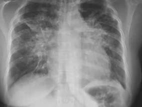
Image 1: Anteroposterior chest radiograph upon admission. Abundant bilateral interstitial infiltrates are observed.
The patient presented a slow clinical progression, characterized by tachypnea and polypnea, requiring oxygen support with a fraction of inspired oxygen (FiO2) of 55 % to achieve oxygen saturations between 89 % and 91 %. On the seventh day of hospitalization, she was transferred to the hospital's Intensive Care Unit, where intravenous ceftriaxone was added to her antimicrobial treatment at a dose of 1 g every 12 hours.
Subsequently, 12 days after admission, the patient experienced retrosternal pain, which led to an electrocardiogram that showed no abnormalities, as well as a repeat chest X-ray in an anteroposterior projection. This study revealed multiple lesions with characteristics of bullous disease and the presence of pneumothorax in both lung fields (Figure 2).
After a re-evaluation of the case, a diagnosis of bilateral bullous disease with a pneumothorax less than 30 % was established. This was interpreted as a manifestation of atypical pneumonia, which contraindicated the use of a pleural drain. It was also considered a contraindication for the implementation of mechanical ventilation.
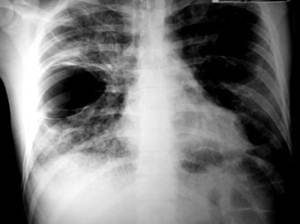
Image 2: AP chest radiograph 15 days after admission. Multiple bullous lesions and pneumothorax in both lung fields were evident.
Given the insufficient clinical response observed on day 15 of hospitalization, vancomycin at a dose of 1 g every 12 hours and amikacin at 550 mg every 12 hours were added to the previously established therapy. During this period, oxygen requirements increased, leading the patient to use a face mask. Subsequent radiographs showed rapid progression in the size of the preexisting bullae, as well as the appearance of new lesions in both hemithoraces. The surgical team reassessed the case and recommended a wait-and-see approach, as placement of an intercostal tube or open surgery could worsen the patient's condition, and he was also unlikely to tolerate such procedures. Although insertion of an airway drainage valve was considered an ideal option, this would only be indicated if the pneumothorax worsened significantly.
The patient remained in critical but stable condition until the 18th day of hospitalization, at which point he experienced an acute episode of severe chest pain on the right side, accompanied by severe respiratory distress, peripheral and perioral cyanosis, and a respiratory rate exceeding 50 breaths per minute. A chest puncture was performed for evacuation purposes; however, this intervention failed to yield significant results, resulting in the patient's death less than an hour after the onset of this crisis due to severe respiratory distress, with no possible recovery from the significant pneumothorax he presented at the time.
It is important to clarify in this case that a lymphocyte count was not available upon admission, nor was it known about his adherence to treatment, as the patient's family stated that she did take them regularly. This suggests another case where antiretroviral therapy is ineffective in some patients, leading to rapid progression to AIDS complications.
DISCUSSION
The disease known until now as "Pneumocystis jirovecii pneumonia" is one of the entities that cause illness and death in people with immunodeficiency. This agent is a pathogen frequently, although not exclusively, associated with advanced stages of HIV infection. It generally causes severe pneumonia in patients with CD4 lymphocyte counts below 200 cells/mm3, with predominantly symptoms of cough, fever, and dyspnea. Radiographic findings most frequently show reticulo-nodular infiltrates, but consolidation or normal lymphocyte counts may also be present. (5)
Pizarro PR et al., (5) in their study explain how the most frequent clinical presentation occurs in AIDS patients with a T-helper lymphocyte (CD4) count below 200 cells/mm3. Common symptoms include progressive dyspnea, nonproductive cough, and low-grade fever. Acute dyspnea with chest pain may indicate a pneumothorax. Physical examination revealed tachypnea, tachycardia, and normal auscultation. Laboratory findings included an elevated LDH, which is highly sensitive in the presence of this germ but not very specific, and is therefore of limited value for the diagnosis of patients with AIDS and pneumonia. These results are consistent with this case, where the patient was admitted with severe respiratory distress, cough, and low-grade fever upon physical examination.
Cantarelli L et al., (6) propose that imaging and carbon monoxide diffusion tests have proven useful. In a prospective study, they found that a normal chest x-ray with no changes or with an uncertain reading ruled out the disease in almost all cases. This data does not correspond to the case presented, due to a nonspecific image with abundant interstitial infiltrates. These radiological images are common in several respiratory infectious pathologies.
Zuluaga I, (7) explains in his research that, in patients with HIV infection, the occurrence of pneumothorax is uncommon, although an increase has been observed in recent years, having an inverse relationship with the number of CD4 lymphocytes, with a frequency that varies between 2,7 % and 4,9 %, much higher than in non-immunocompromised patients. Among patients with HIV infection, this incidence can reach 9 % if P. jirovecii infection coexists; in non-coinfected patients, the frequency is zero, results that are inconsistent with this case, where the patient rapidly progressed to pneumothorax and bullae in both lungs.
Martín PL et al. (8) in their study suggest that HIV-infected patients who develop pneumothorax have a grim prognosis. The impact of the onset of pneumothorax is reflected in an increase in their average length of stay to 9 days versus 5 days in patients without pneumothorax; transfers to an Intensive Care Unit are much more frequent (54 % versus 11 %), and an increase in mortality to 30 % versus 5 % in patients without pneumothorax. These results are consistent with this case, where the patient, after a rapid progression to pneumothorax and bullae, died a few days after admission.
The drug of choice for the treatment of Pneumocystis jirovecii pneumonia (PJP) is trimethoprim/sulfamethoxazole (TMP/SMX) as proposed in the study by Zuluaga I (7). Initially, it is administered intravenously, but it can be given orally (PO) on an outpatient basis in patients with mild to moderate disease, and in those who do not have problems with its absorption. The recommended dose is 15 to 20 mg/kg/day of TMP and 75–100 mg/kg/day of sulfamethoxazole, every 6–8 h, for 21 days in patients with AIDS, and in other patients for 14 days, in this case different antibiotics were used, because there was no certainty of the causal germ, although a broad spectrum antimicrobial therapy was used for its treatment.
In the study by Zuluaga I (7), it is reaffirmed that the use of corticosteroids is necessary in patients with severe clinical symptoms, with oxygen saturation <70 mmHg in arterial blood gases or an alveolar-arterial difference >35 mmHg (AI). They should be started within the first 72 hours of starting the antibiotic, and a regimen of 40 mg of oral prednisone every 12 hours for 5 days is recommended, followed by 40 mg every day for 5 days, and completing 11 days with 20 mg/day. If parenteral administration is required, methylprednisolone can be used at 75 % of the corresponding prednisolone dose. This result corresponds to those proposed in the study by Li H et al. (9) regarding the use of steroids in this serious complication. Tritle BJ et al. (10) argue in their research that despite the use of TMP-SMX as the therapy of choice for both prophylaxis and treatment of PJP, lack of response or intolerance to it has led to the use of pentamidine as a safe and effective alternative in clinical practice.
The therapies indicated for the onset of pneumothorax in a patient with HIV infection are varied, but with poor response, especially those caused by infections, as many present with prolonged bronchopleural fistulas as a result of alterations in the underlying parenchyma. (7)
The use of intercostal tubes connected to a closed underwater system with continuous suction and regulated pressure is the first and most common surgical technique used in these cases, with good results in small, unilateral pneumothoraces without bronchopleural fistulas, as proposed by Alsayed AR et al. (11) in their study. This differs from this case, where the use of an intercostal tube was discouraged due to the small pneumothorax, even though this proved to be a fatal complication in the following days.
Mortality in AIDS patients complicated by pneumonia due to (PJP) ranges between 10 % and 20 % during the initial period of infection, but increases if patients arrive at intensive care units requiring mechanical ventilation, as occurred in our case. Slowly progressive forms of the infection, atypical forms with complications such as pneumothorax, have been reported to be associated with a significant increase in deaths. Mortality is estimated to increase to 75-100 % in patients who do not respond to therapy within the first 5-10 days. (8,10)
CONCLUSIONS
PJP remains an opportunistic disease in immunocompromised patients, especially those with AIDS. Infection with this pathogen in HIV-negative patients is an uncertain battleground; diagnostic techniques and therapeutic approaches are not fully validated. Furthermore, changes in pulmonary physiology and anatomy in this group of patients mean that adjuvant corticosteroid therapy is not preferred. Additional research is needed to determine the best treatment options for these patients to reduce the associated morbidity and mortality and thus allow for a more accurate diagnosis when infection with this pathogen occurs.
BIBLIOGRAPHIC REFERENCES
1. Mocelin HJS, de Jezus SV, Negri LDSA, Borges BJP, da Silva AI, Maciel ELN. Barreiras e facilitadores do enfrentamento de HIV/aids e sífilis por venezuelanas residentes no Brasil. Rev Panam Salud Pública [Internet]. 2023 [cited 09/10/2024]; 3; 47:e3. Available in: https://doi.org/10.26633/RPSP.2023.3
2. Bonnet P, Le Gal S, Calderon E, Delhaes L, Quinio D, Robert-Gangneux F, et al. Pneumocystis jirovecii in Patients With Cystic Fibrosis: A Review. Front Cell Infect Microbiol[Internet]. 2020 [cited 09/10/2024]; 29; 10:571253. Available in: https://doi.org/10.3389/fcimb.2020.571253
3. Solano L MF, Alvarez Lerma F, S. Grau S, Segura C, Aguilar A. Neumonía por pneumocystis jiroveci: características clínicas y factores de riesgo asociados a mortalidad en una unidad de cuidados intensivos. Cuad. - Hosp. Clín [Internet]. 2015 [cited 09/10/2024]; 56(1): 69-69. Available in: http://www.scielo.org.bo/scielo.php?script=sci_arttext&pid=S1652-67762015000100010&lng=es
4. Hernández S, Puerto MP, Gomez C. Neumonía por Pneumocystis Jirovecii en paciente adolescente inmunosuprimido no VIH positivo: Un reporte de caso. Infect [Internet]. 2021 [cited 09/10/2024]; 25(1): 59-62. Available in: http://www.scielo.org.co/scielo.php?script=sci_arttext&pid=S0123-93922021000100059&lng=en
5. Pizarro PR, Valdés HC, Vitali CJ. Presentación inhabitual de un caso de neumonía bulosa aguda bilateral por Pneumocystis jiroveci complicada con neumotórax. Rev. chil. infectol [Internet]. 2007 [cited 09/10/2024]; 24(1): 68-71. Available in: http://www.scielo.cl/scielo.php?script=sci_arttext&pid=S0716-10182007000100012&lng=es
6. Cantarelli L, Gutiérrez Nicolás F, Nazco Casariego GJ, García Gil S. Adecuación a las recomendaciones diagnósticas en pacientes con neumonía por Pneumocystis jirovecii tratados con pentamidina intravenosa. Rev Esp Quimioter[Internet]. 2022 [cited 09/10/2024]; 35(1):30-34. Available in: https://doi.org/10.37201/req/064.2021
7. Zuluaga I. Protocolo de estudio y manejo de infección por Pneumocystis jirovecii, Study protocol and management of Pneumocystis jirovecii infections Rev colom Infectio [Internet]. 2012 [cited 09/10/2024]; 16 (3): 129-131. Available in: https://www.elsevier.es/es-revista-infectio-351-articulo-protocolo-estudio-manejo-infeccion-por-S0123939212700401
8. Martín Pedraz L, Carazo Gallego B, Moreno Pérez D. Clinical-epidemiological characteristics of Pneumocystis jirovecii pneumonia in a tertiary hospital in Spain. An Pediatr (Engl Ed) [Internet]. 2021 [cited 09/10/2024]; 95(1):4-10. Available in: https://doi.org/10.1016/j.anpede.2020.04.023
9. Li H, Lu Y, Tian G, Wu Y, Chen T, Zhang J, et al. A regimen based on the combination of trimethoprim/sulfamethoxazole with caspofungin and corticosteroids as a first-line therapy for patients with severe non-HIV-related pneumocystis jirovecii pneumonia: a retrospective study in a tertiary hospital. BMC Infect Dis [Internet]. 2024 [cited 09/10/2024]; 31; 24(1):152. Available in: https://doi.org/10.1186/s12879-024-09031-7
10. Tritle BJ, Hejazi AA, Timbrook TT. The effectiveness and safety of low dose trimethoprim-sulfamethoxazole for the treatment of pneumocystis pneumonia: A systematic review and meta-analysis. Transpl Infect Dis [Internet]. 2021 [cited 09/10/2024]. Available in: https://doi.org/10.1111/tid.13737
11. Alsayed AR, Al-Dulaimi A, Alkhatib M, Al Maqbali M, Al-Najjar MAA, Al-Rshaidat MMD. A comprehensive clinical guide for Pneumocystis jirovecii pneumonia: a missing therapeutic target in HIV-uninfected patients. Expert Rev Respir Med [Internet]. 2022 [cited 09/10/2024]; 16(11-12):1167-1190. Available in: https://doi.org/10.1080/17476348.2022.2152332
STATEMENT OF AUTHORSHIP
PFAT: Conceptualization, research, methodology, project administration, validation, original draft, review, editing.
FADB: Conceptualization, research, methodology, validation, original draft, review, editing.
CONFLICTS OF INTEREST
The authors declare that there are no conflicts of interest related to this study.
FUNDING
The authors did not receive funding for the development of this study.