CASE PRESENTATION
Acute coronary syndrome without ST-segment elevation secondary to coronary fistula. Case presentation
Síndrome coronario agudo sin elevación del segmento ST secundario a fístula coronaria. Presentación de caso
Hadierkis Sarai Coello Kindelan 1, https://orcid.org/0009-0002-5719-0848
Luis Angel Zayas Massó 2*, https://orcid.org/0009-0000-5397-2776
Andrés Rosell Oliva 3, https://orcid.org/0009-0006-5935-6978
Branly Armando Planas Díaz 2, https://orcid.org/0009-0002-9575-1183
1 University of Medical Sciences of Santiago de Cuba. Faculty of Medicine No. 1. Santiago de Cuba, Cuba.
2 University of Medical Sciences of Santiago de Cuba. Faculty of Medicine No. 2. Santiago de Cuba, Cuba.
3 Saturnino Lora Provincial Hospital. Santiago de Cuba, Cuba.
* Corresponding author:luiszayasmasso@gmail.com
Received: 19/04/2026
Accepted: 08/08/2026
Published: 10/08/2026
How to cite this article: Coello-Kindelan HS; Zayas-Massó LA; Rosell-Oliva A; Planas-Díaz BA. Acute coronary syndrome without ST-segment elevation secondary to coronary fistula. Case presentation. MedEst. [Internet]. 2026 [cited access date]; 6:e541. Available in: https://revmedest.sld.cu/index.php/medest/article/view/541
ABSTRACT
Introduction: Coronary artery fistulas are rare vascular anomalies that may alter myocardial perfusion and mimic acute coronary syndromes. Their clinical relevance lies in the fact that they can occur in the absence of obstructive coronary artery disease, representing a diagnostic challenge.
Objective: To describe a clinical case of non-ST-segment elevation acute coronary syndrome secondary to a coronary artery fistula.
Case presentation: A 53-year-old female patient with a history of arterial hypertension, grade I obesity, and chronic smoking presented with typical ischemic chest pain associated with dyspnea and autonomic symptoms. Electrocardiography showed dynamic ischemic changes with subsequent normalization. Cardiac biomarkers revealed elevated troponin T and creatine kinase MB fraction levels. Transthoracic echocardiography demonstrated regional wall motion abnormalities with preserved left ventricular ejection fraction. Coronary angiography revealed normal epicardial coronary arteries without atherosclerotic lesions and a fistula originating from the left anterior descending artery with drainage into the pulmonary circulation (ICD-11: Q24.5, I20.0). Conservative medical treatment was indicated, with favorable clinical evolution.
Conclusions: Coronary artery fistulas may present as non-ST-segment elevation acute coronary syndrome in the absence of obstructive coronary disease. Coronary angiography is essential for definitive diagnosis and therapeutic decision-making. Conservative management may be effective in hemodynamically stable cases without significant shunt.
Keywords: Acute Coronary Syndrome; Coronary Angiography; Coronary Vessel Anomalies; Myocardial Ischemia; Pulmonary Artery.
RESUMEN
Introducción: Las fístulas de las arterias coronarias son anomalías vasculares infrecuentes que pueden alterar la perfusión miocárdica y simular síndromes coronarios agudos. Su reconocimiento es clínicamente relevante debido a que pueden presentarse sin enfermedad coronaria obstructiva, lo que constituye un reto diagnóstico.
Objetivo: Describir un caso clínico de síndrome coronario agudo sin elevación del segmento ST secundario a fístula coronaria.
Presentación del caso: Se presenta una paciente femenina de 53 años, con antecedentes de hipertensión arterial, obesidad grado I y tabaquismo crónico. Acudió por dolor torácico típico de características isquémicas, asociado a disnea y síntomas vegetativos. El electrocardiograma evidenció cambios dinámicos de isquemia con normalización posterior. Los biomarcadores cardíacos mostraron elevación de troponina T y creatina quinasa fracción MB. El ecocardiograma reveló alteraciones segmentarias de la contractilidad con fracción de eyección conservada. La coronariografía demostró arterias coronarias epicárdicas sin lesiones ateroscleróticas y una fístula originada en la arteria descendente anterior con drenaje hacia la circulación pulmonar (CIE-11: Q24.5, I20.0). Se indicó tratamiento médico conservador con evolución clínica favorable.
Conclusiones: Las fístulas coronarias pueden manifestarse como síndrome coronario agudo sin elevación del ST en ausencia de enfermedad coronaria obstructiva. La coronariografía resulta esencial para el diagnóstico definitivo y la conducta terapéutica. El manejo conservador puede ser efectivo en casos sin repercusión hemodinámica significativa.
Palabras clave: Angiografía coronaria; Fístula arteriovenosa; Isquemia miocárdica; Síndrome coronario agudo; Trompa pulmonar.
INTRODUCTION
Coronary artery fistulas are rare congenital vascular anomalies characterized by an abnormal communication between an epicardial coronary artery and a cardiac chamber or a large vessel, creating a shunt with variable hemodynamic repercussions. (1) Although most patients remain asymptomatic for decades, these anomalies can manifest as myocardial ischemia, heart failure, arrhythmias, or, in exceptional cases, rupture of the fistulous vessel. (2)
Epidemiologically, coronary fistulas are infrequent in the general population, with reported prevalences between 0.05% and 0.25% using conventional coronary angiography, increasing to approximately 0.9% with the use of coronary computed tomography, demonstrating the greater diagnostic sensitivity of current techniques. (3) In the Cuban context, its detection depends on access to technological resources and experience in hemodynamics, with a low incidence reported, even in local series from centers with interventional activity. (4)
Despite their rarity, these anomalies can have significant clinical repercussions when associated with the coronary steal phenomenon, capable of producing myocardial ischemia even in the absence of significant obstructive lesions. (5) In this context, their presentation as non-ST-segment elevation acute coronary syndrome (STEMI) can complicate the differential diagnosis, leading to initial misinterpretations if this entity is not considered among the non-atherosclerotic causes of ischemia. (5,6)
This case is of interest for three main reasons: (i) the coexistence of multiple cardiovascular risk factors in a patient that initially masked a non-atherosclerotic cause, (ii) the demonstration of objective myocardial ischemia with elevated biomarkers and dynamic electrocardiographic abnormalities, in the complete absence of obstructive coronary artery disease, and (iii) the success of conservative management in a low-flow fistula, which reinforces the importance of hemodynamic stratification for therapeutic decision-making.
The scarcity of national reports and the diagnostic complexity inherent in this condition justify the presentation of cases that contribute to the understanding of its clinical behavior and the optimization of its therapeutic management.
The objective of this paper is to describe a clinical case of acute coronary syndrome without ST-segment elevation secondary to a coronary fistula of the left anterior descending artery draining into the pulmonary circulation, diagnosed by coronary angiography and managed conservatively with a favorable outcome.
CASE PRESENTATION
We present the case of a 53-year-old female patient with a long-standing history of hypertension, routinely treated with captopril 6.25 mg daily and hydrochlorothiazide 25 mg daily. She also presented with grade I obesity and a body mass index of 31.2 kg/m², and a smoking history of over 20 years.
She went to the emergency department of the Saturnino Lora Provincial Clinical Surgical Teaching Hospital in Santiago de Cuba, with sudden onset of intense, oppressive, and burning precordial pain radiating to her neck, jaw, and left upper extremity, lasting more than 30 minutes and unrelieved by rest. The symptoms were accompanied by exertional dyspnea, cold sweats, pallor, nausea, and a generalized feeling of weakness. The patient reported no previous similar episodes.
On physical examination, increased subcutaneous fat was noted. Cardiovascular examination revealed tachycardia (heart rate of 102 beats per minute), muffled heart sounds without murmurs, and delayed capillary refill (greater than 3 seconds). Peripheral vascular examination showed weak and rapid arterial pulses, as well as pitting edema in both lower extremities, extending to the lower third of the legs. The edema was cold, non-tender, and pitted. Blood pressure was 148/92 mmHg. Oxygen saturation by pulse oximetry was 94% on room air.
A 12-lead electrocardiogram was performed upon admission, which showed abnormalities consistent with myocardial ischemia, characterized by transient ST-segment elevation in the right precordial leads (V1-V3) and negative T waves in the anterolateral leads (I, aVL, V4-V6). A second electrocardiogram, performed 20 minutes later, showed normalization of the ST segment (Figure 1)
Figure 1. 12-lead electrocardiogram performed on admission, showing ventricular repolarization abnormalities characterized by negative T waves in precordial leads and transient ST segment changes.
A cardiac biomarker profile was ordered, which revealed elevated high-sensitivity troponin T (45 ng/L; reference range: <14 ng/L) and creatine kinase MB (CK-MB) (65 U/L; reference range: <25 U/L). The complete blood count showed leukocytosis (12 × 10⁹/L), an elevated erythrocyte sedimentation rate (28 mm/h), and elevated C-reactive protein (5.0 mg/L). Serum creatinine and electrolyte levels were normal.
The transthoracic echocardiogram showed normal-sized cardiac chambers, preserved global systolic function with a left ventricular ejection fraction of 65%, and segmental contractility abnormalities in the territory of the left anterior descending artery (apical and anterior wall hypokinesis). No vegetations, intracardiac masses, or significant pericardial effusion were observed.
Based on these findings, a diagnosis of non-ST-segment elevation acute coronary syndrome (STEMI) was established, and the patient was transferred to the cardiac catheterization laboratory for invasive studies.
Diagnostic coronary angiography was performed via a right radial approach, which showed epicardial coronary arteries without significant atherosclerotic lesions. A coronary fistula originating from the proximal segment of the left anterior descending artery was identified, draining into the right pulmonary circulation, with an approximate diameter of 2.5 mm. The pulmonary-to-systemic flow ratio (Qp/Qs) was less than 1.5. The right coronary artery was found to be patent in all its segments, with no evidence of dissection, thrombosis, or embolization. (Figure 2)
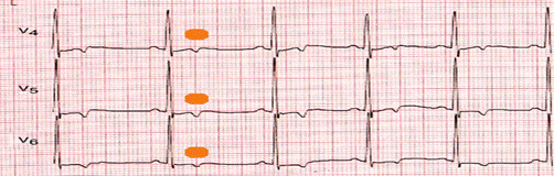
Figure 2. Selective coronary angiography showing the presence of a coronary fistula originating in the proximal segment of the left anterior descending artery with drainage to the right pulmonary circulation, without evidence of atherosclerotic lesions in the epicardial coronary arteries.
The final diagnosis was acute coronary syndrome without ST-segment elevation secondary to a coronary fistula of the left anterior descending artery draining into the pulmonary circulation (ICD-11: Q24.5, I20.0).
Pharmacological treatment was initiated with metoprolol 50 mg every 12 hours, aspirin 125 mg daily, clopidogrel 75 mg daily, enalapril 10 mg every 12 hours, and atorvastatin 40 mg daily, with an adequate clinical response. Percutaneous intervention or surgical closure of the fistula was not indicated due to the absence of significant hemodynamic compromise (Qp/Qs < 1.5), hemodynamic stability, and the absence of signs of heart failure.
During her hospital stay, the patient showed progressive clinical improvement, with resolution of chest pain 8 hours after the start of treatment, hemodynamic stability, and progressive normalization of cardiac biomarkers (troponin T decreased to 18 ng/L by the second day). She remained asymptomatic for the remainder of her hospital stay, with no new electrocardiographic abnormalities or documented arrhythmias. She experienced no hemorrhagic complications or adverse events related to treatment.
She was discharged on the third day of admission with scheduled follow-up appointments at the cardiology outpatient clinic at 2 and 4 weeks, instructions for lifestyle modifications (low-sodium diet, moderate physical activity), permanent cessation of smoking, and strict adherence to the prescribed medical treatment.
Informed consent and ethics: Written informed consent was obtained from the patient for the publication of this clinical case and the associated diagnostic images. Confidentiality of information and anonymity of personal data were guaranteed. The study was conducted in accordance with the ethical principles established in the Declaration of Helsinki for research involving human subjects.
DISCUSSION
The presentation of an acute coronary syndrome without ST-segment elevation in a middle-aged patient with multiple cardiovascular risk factors (hypertension, obesity, and smoking) initially suggested an atherothrombotic etiology. However, the absence of obstructive coronary lesions on coronary angiography and the finding of a coronary fistula with pulmonary drainage constituted an unexpected diagnosis that required a therapeutic reassessment. This scenario exemplifies the importance of considering non-atherosclerotic causes of myocardial ischemia in the differential diagnosis of ST-segment elevation myocardial infarction (STEMI), particularly when the angiographic findings do not correlate with the clinical presentation. (1,5)
From a pathophysiological perspective, coronary fistulas create an arteriovenous shunt that can produce myocardial ischemia through the phenomenon of "coronary steal." In this mechanism, a significant portion of blood flow is diverted to the low-resistance territory (in this case, the pulmonary circulation), reducing distal perfusion of the affected coronary territory. (5)
Torres et al. (5) have demonstrated that this phenomenon can mimic a type 1 acute myocardial infarction, even when the underlying mechanism is functional and not obstructive. The location of the fistula in the proximal segment of the left anterior descending artery, as in the case presented, confers greater clinical relevance since this territory supplies blood to a large myocardial mass, increasing the risk of significant hemodynamic compromise. (7)
The initial differential diagnosis for this patient included acute atherothrombotic coronary syndrome, aortic dissection, pulmonary embolism, acute myocarditis, and Takotsubo syndrome. The absence of radiating back pain, normal D-dimer levels, and preservation of global systolic function with focal segmental involvement pointed toward a coronary etiology. However, it was coronary angiography that definitively ruled out obstructive atherosclerotic disease and identified the fistula as the cause of the ischemic event. (2) This finding underscores that, in patients with ST-segment elevation myocardial infarction (STEMI) and epicardial coronary arteries free of atherosclerosis, the presence of congenital coronary anomalies, including fistulas, should be systematically suspected. (6)
The dynamic electrocardiographic changes observed in this patient—transient ST-segment elevation followed by normalization—are consistent with an intermittent ischemic event. Salazar (8) has described that coronary fistulas with pulmonary drainage can produce fluctuating episodes of ischemia, depending on hemodynamic variations and the degree of flow diversion. Rusali et al. (3) indicate that this intermittent electrical behavior is characteristic of fistulas with variable flow, especially in the absence of associated coronary obstruction, which coincides with the pattern observed in this case. The presence of negative T waves in the anterolateral leads also suggested involvement of the left anterior descending artery territory, a finding that was subsequently confirmed by echocardiography and coronary angiography.
Transthoracic echocardiography is an accessible diagnostic tool that, in this case, allowed the detection of segmental contractility disorders in the territory of the left anterior descending artery with preserved global systolic function. This pattern, also described by Acitelli et al. (6) In patients with coronary fistulas diagnosed during an ST-segment elevation myocardial infarction (STEMI), this reflects regional hypoperfusion secondary to the diversion of blood flow, without necessarily implying extensive myocardial necrosis. The preserved ejection fraction of 65% in this patient, while reassuring, did not exclude the risk of recurrent ischemic events, which is why strict medical management and close monitoring were chosen.
The decision to manage conservatively in this patient was based on three fundamental criteria: (a) hemodynamic stability from admission; (b) absence of significant hemodynamic compromise, evidenced by a Qp/Qs ratio of less than 1.5; and (c) a fistula diameter of less than 3 mm, the generally accepted threshold for considering closure in the absence of severe symptoms. (4,9)
Araújo-Leite et al. (4) and Favaloro et al. (9) agree that percutaneous or surgical closure should be reserved for high-flow fistulas (Qp/Qs > 1.5), symptomatic fistulas refractory to medical treatment, or those associated with complications such as endocarditis, thrombosis, or heart failure. In this context, pharmacological management with beta-blockers, dual antiplatelet agents, angiotensin-converting enzyme inhibitors, and statins, along with strict modification of risk factors, constituted an appropriate strategy that resulted in a favorable outcome. Soledispa et al. (7) described a coronary-pulmonary fistula as the cause of acute coronary syndrome in an adult patient, with comparable angiographic findings and clinical course. However, unlike that report, in which percutaneous closure was chosen, the present case justified conservative management due to the absence of high flow and clinical stability.
The positive clinical response to conservative treatment, with pain resolution within 8 hours and a progressive decrease in myocardial damage biomarkers, is consistent with the results reported by Santiago-Peña et al. (10) in selected patients managed non-invasively. However, it is imperative to emphasize that these conditions require long-term monitoring. Díaz-Sarasa et al. (2) warn of the potential risk of late complications, including shunt progression, development of heart failure, ventricular arrhythmias, and, in exceptional cases, fistula rupture. Therefore, regular outpatient follow-up with echocardiographic evaluation and, if necessary, advanced imaging studies is essential. (2,10)
Lessons learned from this case:
Diagnostic masking: The presence of multiple cardiovascular risk factors (hypertension, obesity, smoking) can divert attention toward an atherothrombotic etiology, even when the underlying mechanism is a congenital anomaly. A normal coronary angiography in a patient with ST-segment elevation myocardial infarction (STEMI) should raise suspicion of non-atherosclerotic causes.
Hemodynamic stratification: Measuring the Qp/Qs ratio and assessing the fistula diameter are essential for deciding between conservative and interventional management. Not all symptomatic fistulas require percutaneous or surgical closure.
Long-term follow-up: Despite a favorable initial outcome, coronary fistulas require prolonged monitoring due to the risk of late complications.
Multidisciplinary approach: Successful management required collaboration between clinical cardiology, interventional cardiology, and the emergency department team, highlighting the importance of teamwork.
This report has inherent limitations in its design. First, it is a single case, which prevents generalizing the findings to broader populations. Secondly, advanced imaging studies such as coronary computed tomography or cardiac magnetic resonance imaging were unavailable, which would have allowed for better anatomical characterization of the fistula and more precise quantification of the shunt flow. Thirdly, post-discharge follow-up was brief (at the time of writing), so it is not possible to evaluate the medium- and long-term evolution or the possible development of late complications. Finally, right heart catheterization and direct measurement of pulmonary pressures were not performed, which would have enriched the hemodynamic characterization.
CONCLUSIONS
We present a case of acute coronary syndrome without ST-segment elevation secondary to a coronary fistula of the left anterior descending artery draining into the pulmonary circulation, diagnosed by coronary angiography. Its non-atherosclerotic nature as the cause of myocardial ischemia was confirmed. Conservative medical treatment showed a favorable outcome without complications. This case highlights the importance of including this entity in the differential diagnosis of acute coronary syndrome without ST-segment elevation in the absence of obstructive coronary artery disease.
BIBLIOGRAPHIC REFERENCES
1- Crespo Valdés J, Muiños Martínez C. Fístula de arteria coronaria descendente anterior a arteria pulmonar en paciente no pediátrico. Reporte de un caso en Cuba. Rev Cuba Cardiol Cir Cardiovasc [Internet]. 2021 [cited 13/04/2026];27(2):e1015. Available in: https://revcardiologia.sld.cu/index.php/revcardiologia/article/view/1015
2- Díaz-Sarasa PD, Méndez-Sánchez HG, Eid-Lidt G. Aneurismas y fístula de arteria coronaria posterior a biopsia endomiocárdica en un paciente con trasplante de corazón. Evolución angiográfica y clínica. Arch Cardiol Mex [Internet]. 2025 [cited 13/04/2026];95(1):111-114. Available in: http://www.scielo.org.mx/scielo.php?script=sci_arttext&pid=S140599402025000100111
3- Santiago-Peña JJ, Saaibi-Solano JF, Rubio-Duarte AF, Pinto-Martínez IA, Molina-Mora Y, Díaz-Caraballo MI. Cierre endovascular de fístula coronaria en el lactante. Arch Cardiol Mex [Internet]. 2024 [cited 13/04/2026];94(4):515-518. Available in: http://www.scielo.org.mx/scielo.php?script=sci_arttext&pid=S140599402024000400515
4- Rusali AC, Lupu CI, Manolache MM, et al. Fístula coronaria-pulmonar en un paciente con dolor torácico atípico: presentación de caso y revisión de la literatura. Surg Radiol Anat. [Internet] 2025 [cited 13/04/2026];47:240. Available in: https://doi.org/10.1007/s00276-025-03724-7
5- Araújo-Leite MP, Costa J, Galvão-Braga C. Tratamiento de fístula arterial coronaria iatrogénica. REC Interv Cardiol ES [Internet]. 2023 [cited 13/04/2026];5(1):83-84. Available in: http://scielo.isciii.es/scielo.php?script=sci_arttext&pid=S260473062023000100018
6- Torres C, Gjergjindreaj M, Torres-Ortiz H, Fuentes J, Beohar N. Coronary steal syndrome secondary to large coronary to pulmonary artery fistulas. Cureus. [Internet] 2022 [cited 13/04/2026];14(10):e30267. Available in: https://doi.org/10.7759/cureus.30267
7- Acitelli A, Bencivenga S, Giannico MB, et al. Coronary artery fistula diagnosed by echocardiography during NSTEMI: case report and review of literature. Case Rep Cardiol. [Internet] 2019 [cited 13/04/2026]; 2019:5956806. Available in: https://doi.org/10.1155/2019/5956806
8- Soledispa CI, Larrea M, Alberca SS, Zelaya A, Sanango K. Fístula coronaria-pulmonar como causa de síndrome coronario agudo. Rev Argent Cardiol [Internet]. 2023 [cited 13/04/2026];91(2):159-160. Available in: https://www.scielo.org.ar/scielo.php?script=sci_arttext&pid=S185037482023000200159
9- Salazar FJ. Fístulas coronario-pulmonares asintomáticas. Reporte de caso. Rev Urug Med Int [Internet]. 2020 [cited 13/04/2026];5(3):19-25. Available in: http://www.scielo.edu.uy/scielo.php?script=sci_arttext&pid=S2393-67972020000300019
10- Favaloro RR, Casabé JH, Escarain MC, Ganum GE, Mendiz O. Fístula coronaria gigante resuelta quirúrgicamente. Medicina (B Aires) [Internet]. 2020 [cited 13/04/2026];80(1):91-91. Available in: https://www.scielo.org.ar/scielo.php?script=sci_arttext&pid=S0025-76802020000100013
CONFLICT OF INTEREST
The authors declare no conflict of interest.
AUTHORSHIP CONTRIBUTION
HACK: conceptualization, data curation, formal analysis, research, methodology, drafting of the original manuscript, revision writing, and editing.
LAZM: conceptualization, data curation, formal analysis, research, methodology, drafting of the original manuscript, revision writing, and editing.
ARO: research, methodology, drafting of the original manuscript, revision writing, and editing.
BAPD: research, methodology, drafting of the original manuscript, revision writing, and editing.
FUNDING SOURCES
The authors declare that no external funding was received.
USE OF ARTIFICIAL INTELLIGENCE
The authors declare that generative artificial intelligence was not used in the writing of the manuscript.
This case presentation was prepared following the recommendations of the CARE (Case Report) guidelines.