CASE PRESENTATION
Giant kerion of Celsus in a pediatric patient. Case report
Querion de Celso Gigante en paciente pediátrico. Informe de caso
Alvaro Rojas Mora 1*, https://orcid.org/0000-0001-6805-5364
Pilar María Acuña Aguilarte 2, https://orcid.org/0000-001-6944-7597
1 Calixto García General University Hospital. La Habana, Cuba.
2 Central Havana Pediatric Hospital. La Habana, Cuba.
* Corresponding author: rojasmoraalvaro@gmail.com
Received: 05/05/2026
Accepted: 08/08/2026
Published: 10/08/2026
How to cite this article: Rojas-Mora A; Acuña-Aguilarte PM. Giant kerion of Celsus in a pediatric patient. Case report. MedEst. [Internet]. 2026 [cited access date]; 6:e547. Available in: https://revmedest.sld.cu/index.php/medest/article/view/547
ABSTRACT
Introduction: Kerion of Celsus is a severe inflammatory form of tinea capitis, uncommon in pediatrics, which presents a diagnostic and therapeutic challenge. Its presentation as a giant lesion is exceptional.
Objective: To report a clinical case of giant kerion of Celsus in a pediatric patient and its multimodal management.
Case presentation: A 7-year-old female patient, with no relevant medical history, presented with a scalp lesion of 15 days' duration that did not respond to amoxicillin. Physical examination revealed a 12 x 10 cm reddish-purple tumor-like plaque in the central region, with short, brittle hairs, multiple confluent pustules draining seropurulent exudate (honeycomb sign), painful to palpation, and with inflammatory lymphadenopathy. Direct examination with KOH was positive, and mycological culture identified Trichophyton tonsurans. Treatment was initiated with terbinafine (125 mg/day for 8 weeks), prednisone (25 mg/day with gradual tapering), and cotrimoxazole (12.5 mL every 12 hours for 7 days). The outcome was favorable, with complete resolution, no adverse effects, normal liver enzymes, and full hair regrowth within three months.
Conclusions: Giant kerion of Celsus caused by Trichophyton tonsurans requires early diagnosis and aggressive multimodal treatment. Triple therapy (antifungal + corticosteroid + antibiotic) proved effective and safe, achieving clinical resolution and preventing scarring alopecia. This case has significant educational value.
Keywords: Kerion of Celsus; Tinea capitis; Trichophyton tonsurans; Pediatrics; Terbinafine.
RESUMEN
Introducción: El Querion de Celso es una forma inflamatoria grave de la tiña capitis, poco frecuente en pediatría, que representa un reto diagnóstico y terapéutico. Su presentación como lesión gigante es excepcional.
Objetivo: Reportar un caso clínico de Querion de Celso Gigante en una paciente pediátrica y su manejo multimodal.
Presentación del caso: Paciente femenina de 7 años, sin antecedentes relevantes, con lesión en cuero cabelludo de 15 días de evolución que no respondió a amoxicilina. Al examen físico se evidenció una placa tumoral rojizo-violácea de 12 x 10 cm en región central, con pelos cortos y quebradizos, múltiples pústulas confluentes que drenaban exudado seropurulento (signo del panal de abeja), dolorosa a la palpación y con adenopatías inflamatorias. El examen directo con KOH fue positivo y el cultivo micológico identificó Trichophyton tonsurans. Se indicó tratamiento con terbinafina (125 mg/día por 8 semanas), prednisona (25 mg/día con reducción gradual) y cotrimoxazol (12.5 mL cada 12 horas por 7 días). La evolución fue favorable, con resolución completa, ausencia de efectos adversos, enzimas hepáticas normales y rebrote capilar total a los tres meses.
Conclusiones: El Querion de Celso gigante por Trichophyton tonsurans requiere diagnóstico temprano y tratamiento multimodal agresivo. La triple terapia (antifúngico + corticoide + antibiótico) resultó eficaz y segura, logrando resolución clínica y previniendo la alopecia cicatricial. Este caso posee un alto valor didáctico.
Palabras clave: Querion de Celso; Tiña capitis; Trichophyton tonsurans; Pediatría; Terbinafina.
INTRODUCTION
Kerion of Celsus is a severe inflammatory form of tinea capitis, uncommon in pediatrics, characterized by a painful, suppurative, tumorous plaque with alopecia and regional lymphadenopathy. It constitutes between 5 % and 10 % of tinea capitis cases, predominantly affecting prepubertal children (1,2,3).
The most common causative agents are Trichophyton and Microsporum, and the diagnosis is confirmed by direct examination with KOH and mycological culture (4). Treatment requires systemic antifungal therapy (terbinafine or griseofulvin), and oral corticosteroids may be added in highly inflammatory cases, along with antibiotics for bacterial superinfection (5).
Most kerion of Celsus described in the literature are between 3 and 8 cm in diameter. Lefranc et al. (6) reported a 10 cm lesion in an adolescent, which they considered extensive and severe. However, the presentation of a giant lesion (greater than 10 cm) is exceptional and poorly documented.
This case is of particular clinical and educational relevance because it documents a 12 x 10 cm kerion, which represents the maximum expression of this condition, and also illustrates a successful multimodal management approach that prevented scarring alopecia.
The objective of this report is to describe a case of giant kerion of Celsus caused by Trichophyton tonsurans in a 7-year-old pediatric patient, along with its corresponding diagnostic and therapeutic approach. The case has been written following the CARE guidelines for case presentations.
CASE PRESENTATION
A 7-year-old female patient, school-aged, with no significant past medical history or known drug allergies, presented to the Dermatology Service of the Central Havana Pediatric Hospital accompanied by her mother, who reported a scalp lesion that had been present for approximately 15 days. Initially, the patient was evaluated by her family physician, who prescribed amoxicillin. However, the lesion did not improve; instead, it worsened, increasing in size, exuding, and becoming painful.
Dermatological Physical Examination
A well-defined, circular, soft, tumor-like plaque was observed in the central region of the scalp. It measured 12 cm along its longitudinal axis and 10 cm along its transverse axis, and was erythematous-violaceous in color. The surface of the lesion was partially devoid of hair, with only short, brittle, and dystrophic-appearing hairs visible. Multiple pustules of varying sizes emerge from the lesion, merging together and draining seropurulent exudate, forming the characteristic honeycomb sign typical of kerion of Celsus (Figure 1). The lesion is painful to the touch and is accompanied by inflammatory lymphadenopathy in the retroauricular and occipital regions.
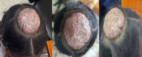
Figure 1: A well-defined, circular, 12 x 10 cm, erythematous-violaceous tumorous plaque was found in the central region of the scalp, with a partially hairless surface. Short, brittle, and dystrophic hairs emerged from it, along with multiple confluent pustules draining seropurulent exudate, forming the honeycomb pattern characteristic of Kerion of Celsus..
Supplementary Examinations:
Direct examination with potassium hydroxide (20% KOH): Septate hyaline hyphae and arthroconidia arranged inside and around the hairs (ectotrix pattern) were observed, compatible with dermatophytes of the genera Microsporum or Trichophyton.
Mycological culture on Sabouraud agar with chloramphenicol and cycloheximide: Positive for Trichophyton tonsurans.
Wood's lamp: Negative, without fluorescence.
Bacterial culture of purulent exudate: Staphylococcus aureus sensitive to methicillin, clindamycin, trimethoprim-sulfamethoxazole and cefixime was isolated.
Complete blood count and erythrocyte sedimentation rate (ESR): Moderate leukocytosis (12,500 cells/mm³) with neutrophilia (78%) and elevated ESR (32 mm/h).
Liver enzymes (pre-antifungal treatment evaluation): ALT (TGP): 28 U/L, AST (TGO): 26 U/L, GGT: 18 U/L (all within normal ranges for age).
Treatment:
Topical treatment: Rosemary (Rosmarinus officinalis) promotions: Local application of compresses soaked in the infusion of the plant (fresh or dried leaves in boiling water, left to warm) on the injury, twice a day, for 10-15 minutes. It is used for its antiseptic, anti-inflammatory and healing properties, as an adjuvant in the management of inflammation and exudation.
Systemic Treatment
Oral antifungal (etiological therapy): Terbinafine 5 mg/kg/day. Calculation: 25 kg × 5 mg/kg = 125 mg/day. Presentation: 250 mg tablets. ½ tablet (125 mg) was administered once a day, orally, after a meal, for 8 weeks.
Oral corticosteroid (anti-inflammatory effect): Prednisone 1 mg/kg/day. Calculation: 25 kg × 1 mg/kg = 25 mg/day. Presentation: 5 mg tablets. 5 tablets (25 mg) were administered once daily in the morning, with food, with gradual reduction: days 1 to 7: 25 mg/day; days 8 to 10: 12.5 mg/day; days 11 to 14: 5 mg/day, then suspension.
Oral antibiotic (bacterial superinfection): Trimethoprim/sulfamethoxazole (cotrimoxazole) in pediatric suspension (40 mg trimethoprim + 200 mg sulfamethoxazole per 5 ml). 12.5 ml was administered orally every 12 hours for 7 days.
Complementary local measures: Gentle cleaning of the lesion with sterile saline solution and gauze was indicated, twice a day, to eliminate exudate and scabs, without applying topical antifungal products or antibiotics.
Evolution and monitoring
The patient was re-evaluated 7, 14, 30 and 60 days after starting treatment in the dermatology outpatient clinic of the Pediatric Hospital of Central Havana. In the first week, a notable improvement in pain, swelling and exudation was observed, with the pustules beginning to dry out. At 14 days, the swelling had almost completely subsided, allowing prednisone to be discontinued as scheduled. After a month of treatment, the tumor plaque had shrunk to less than half its original size (5 x 4 cm), with evident regrowth of healthy hair at the edges.
Liver enzymes (ALT, AST, GGT) were monitored, which remained within normal ranges, with no evidence of hepatotoxicity due to terbinafine. After 8 weeks, the terbinafine cycle was completed, achieving complete clinical resolution of the condition; liver enzymes were repeated, remaining normal. At the 3-month follow-up, complete hair growth was observed in the affected area, with no scar or signs of recurrence. No complications were recorded during the entire treatment and follow-up period.
Informed consent: Written informed consent was obtained from the patient's mother for the publication of this clinical case, guaranteeing the confidentiality of her identity by eliminating identifiable data.
DISCUSSION
Kerion of Celsus is a severe inflammatory form of tinea capitis, uncommon in daily clinical practice, which presents a diagnostic and therapeutic challenge in pediatrics (1-3). Most published cases describe lesions between 3 and 8 cm in diameter. Lefranc et al. (6) reported a 10 cm lesion in an adolescent, considering it extensive and severe. In contrast, the patient presented in this paper showed a 12 x 10 cm plaque, which surpasses even that case and constitutes, to the best of our knowledge, one of the largest lesions documented in the literature. This exceptional size confers high didactic and clinical value.
The patient's clinical presentation (erythematous-violaceous tumor-like plaque, confluent pustules with seropurulent exudate forming a honeycomb pattern, partial alopecia, and painful satellite lymphadenopathy) fully coincides with the classic descriptions of the condition (4,9). Unlike other reports where kerion is associated with dermatophytid reactions or erythema nodosum (5,6), the patient did not present with distant cutaneous manifestations, which simplified the diagnostic approach.
Mycological culture identified Trichophyton tonsurans, an agent that, according to multiple authors, is increasingly common in inflammatory forms of tinea capitis in urban areas (2,11,15,16). Negative fluorescence under Wood's lamp was consistent with this etiology, unlike cases caused by Microsporum spp., which are usually fluorescent (13,17,18).
The patient received terbinafine (125 mg/day for 8 weeks), a drug currently considered first-line for tinea capitis caused by Trichophyton tonsurans (7,11,14,16). Griseofulvin, although historically the standard treatment, is known to require longer courses of treatment and presents a higher risk of interactions (20). The choice of terbinafine for 8 weeks achieved complete clinical resolution without adverse effects or elevated liver enzymes, supporting its safety in pediatrics.
The use of prednisone (1 mg/kg/day with gradual tapering) in kerion is controversial. However, several authors explicitly recommend corticosteroids in moderate to severe cases to reduce inflammation, alleviate pain, and prevent cosmetic sequelae (1,4,8). The patient experienced no adverse effects and achieved resolution without scarring, supporting the indication of corticosteroids in giant kerions. Al Aboud and Crane (14) prefer to avoid them due to a theoretical fear of immunosuppression, but acknowledge that there is no solid evidence to support this concern in short-course regimens.
Completing the treatment regimen, cotrimoxazole was administered for 7 days due to the isolation of susceptible Staphylococcus aureus in the purulent exudate. The need for antibiotics in kerion depends on the presence of bacterial superinfection. Cedeño Roballino et al. (10) and Krize-Morun et al. (12) recommend oral antibiotics when there is clinical or microbiological evidence of superinfection, especially in highly exudative lesions or those with intense pain. In the patient presented here, the presence of abundant purulent exudate and the isolation of S. aureus fully justified their use. Not all authors recommend routine antibiotics (11), but in extensive or complicated cases, triple therapy (antifungal + corticosteroid + antibiotic) has proven effective (10,12).
The patient's favorable outcome, without complications and with complete hair regrowth at three months, is similar to that described in most pediatric cases (8,9,12). No adverse drug reactions or recurrence were recorded, which contrasts with some reports where dermatophytid reactions or drug eruptions were observed (2,19).
In conclusion, giant kerion of Celsus caused by Trichophyton tonsurans in school-aged children requires a timely diagnostic approach (direct examination, mycological culture, and Wood's lamp examination) and combined therapeutic approach (oral antifungal, corticosteroid, and antibiotic when superinfection is present) to ensure complete resolution and prevent aesthetic sequelae. The case presented is a successful example of this multimodal management.
CONCLUSIONS
Giant kerion of Celsus, although infrequent in daily dermatological practice, is a severe form of tinea capitis that requires early diagnosis and timely treatment to avoid irreversible aesthetic sequelae. This case is particularly valuable for two reasons: the giant size of the lesion (12 x 10 cm) and the excellent clinical response obtained with a triple therapeutic regimen combining terbinafine, prednisone, and cotrimoxazole. The favorable evolution, without adverse effects, with normal liver enzymes and complete hair regrowth at three months, confirms the efficacy and safety of this therapeutic strategy.
BIBLIOGRAPHIC REFERENCES
1. Herrera Buitrago MC, Corredor López KS, Fonseca López FF, Rodríguez Estupiñán AC. Querion de Celso moderado, un reto diagnóstico y terapéutico en la infancia. Rev Hispano-Am Dermatol Pediátr. 2025;2(2). doi:10.29176/30284163.2043.
2. Yasuda-Sekiguchi F, Kamata A, Hosokawa R, Kouno M, Takahashi S, Yaguchi T, et al. A Case of Kerion Celsi Caused by Trichophyton tonsurans, a Plate Culture of Which Showed Yellow-Green Fluorescence Under UVA Light. Med Mycol J. 2022;63(2):37-41. doi:10.3314/mmj.21-00022.
3. Chaves Chavarría MN, Molina Cruz T. Querión de Celso, manejo de la dermatofitosis en el cuero cabelludo en el primer nivel de atención. Rev Electron Portalesmedicos [Internet]. 2024 [cited 20/04/2026];19(16):671. Available in: https://www.revista-portalesmedicos.com/revista-medica/querion-de-celso-manejo-de-la-dermatofitosis-en-el-cuero-cabelludo-en-el-primer-nivel-de-atencion/
4. 1. Rivero-Bermúdez MD, Andazora-González EC, Vivas Toro SC. Tiña inflamatoria querion de Celso presentación de un caso. Rev Asoc Colomb Dermatol. 2024;32(3):251-256. doi:10.29176/30284163.2043.
5. Lefranc M, Accoceberry I, Milpied B, Doutre MS, Seneschal J, Boniface K. Severe kerion Celsi caused by Trichophyton quinckeanum. J Mycol Med. 2024;34(1):101453. doi:10.1016/j.mycmed.2023.101453
6. Herzum A, Garibeh E, Gariazzo L, Occella C, Viglizzo G. Erythema nodosum triggered by kerion celsi in pediatrics: literature review and case report. An Bras Dermatol. 2024;99(2):312-315. doi:10.1016/j.abd.2023.03.008
7. Aguinaga Inurriaga AE, Lopez Gutierrez AF. Querión de Celso en el embarazo tratado con terbinafina. Dermatol Rev Mex. 2024;68(2):214-219. doi:10.24245/dermatolrevmex.v68i2.10241
8. Chiriac A, Diaconeasa A, Voicu C, Ivaniciuc M, Miulescu R, Chiriac AE, et al. Kerion Celsi in infants and children - A narrative review 2010-2023. Mycoses. 2024;67(1):e13675. doi:10.1111/myc.13675
9. Montiel Centurión JN, Mongelós Ortiz AB, Da Ponte MN, Ibáñez ME, Aldama Caballero AB. Querión de Celso: a propósito de un caso. Rev Nac (Itauguá). 2024;16(2):155-162. doi:10.18004/rdn2024.may.02.155.162
10. Cedeño Roballino ZS, Tayupanta Alban MA, Gallegos Polo JN, Chávez Almeida JF, Muñoz Ponce AL, Soria Frias PK, et al. El querion de Celso: estrategias actuales en diagnóstico y tratamiento. Braz J Implantol Health Sci. 2024;6(4):47-56. doi.org/10.36557/2674-8169.2024v6n4p47-56
11. Handler MZ, Elston DM. Tinea capitis treatment & management. In: Medscape. New York (NY): WebMD LLC; [Internet] 2024 [cited 20/04/2026]. Available in: https://emedicine.medscape.com/article/1091351-treatment
12. Krize-Morun I, Matamoros-Montoya K, Segnini-Zamora A, Molina-Brays C, Bogantes-Barrantes C. Querión Celso, dermatofitosis del cuero cabelludo infantil. Acta Méd Costarric. 2023;65(1):32-36. doi:10.51481/amc.v65i1.1276
13. Shimoyama H, Taira H, Satoh K, Tamura T, Yo A, Sei Y, et al. Kerion Celsi due to Microsporum canis in an adult woman, treated successfully with fosravuconazole. Med Mycol J. 2023;64(2):37-43. doi:10.3314/mmj.22-00024
14. Al Aboud A, Crane J. Tinea capitis. In: StatPearls Treasure Island (FL): StatPearls Publishing; [Internet] 2023 [cited 20/04/2026]. Available in: https://www.ncbi.nlm.nih.gov/books/NBK536909/
15. Yasuda-Sekiguchi F, Kamata A, Hosokawa R, Kouno M, Takahashi S, Yaguchi T, et al. A case of kerion celsi caused by Trichophyton tonsurans, a plate culture of which showed yellow-green fluorescence under UVA light. Med Mycol J. 2022;63(2):37-41. doi:10.3314/mmj.22-00001
16. Aguirre Sotelo JP, Tarango Martinez VM, Vera Cabrera L. Kerion celsi caused by Trichophyton tonsurans in an adult. An Bras Dermatol. 2022;97(5):637-640. doi:10.1016/j.abd.2021.09.016
17. Chiriac A, Birsan C, Mares M, Wollina U. Kerion Celsi due to Microsporum canis infection. Hautarzt. 2021;72(10):855-859. doi:10.1007/s00105-021-04874-0
18. Deh A, Diongue K, Diadie S, Diatta B, Diop K, Ndour N, et al. Kerion celsi due to Microsporum audouinii: a severe form in an immunocompetent girl. SAGE Open Med Case Rep. 2021;9:2050313X211020486. doi:10.1177/2050313X211020486
19. Nenoff P, Manos A, Ehrhard I, Krüger C, Paasch U, Helmbold P, et al. Kerion Celsi due to Trichophyton soudanense and Pityriasis rosea following treatment by terbinafine. J Dtsch Dermatol Ges. 2021;19(7):1048-1051. doi:10.1111/ddg.14432
20. Leung AKC, Hon KL, Leong KF, Barankin B, Lam JM. Tinea capitis: an updated review. Recent Pat Inflamm Allergy Drug Discov. 2020;14(1):58-68. doi:10.2174/1872213X14666200106145624
AUTHORSHIP CONTRIBUTION
ARM: Conceptualization, research, data curation, methodology, visualization, drafting of the original manuscript, and revision and editing of the final manuscript.
PMAG: Conceptualization, research, and supervision.
CONFLICT OF INTEREST
The authors declare no conflict of interest.
FUNDING SOURCES
The authors received no funding for the development of this article.
USE OF ARTIFICIAL INTELLIGENCE
The authors declare that no artificial intelligence was used in the preparation of this manuscript.
This case presentation was prepared following the recommendations of the CARE (Case Report) guidelines.