CASE PRESENTATIONS
Congenital telangiectatic cutis marmorata: a case report
Cutis marmorata telangiectásico congénito: a propósito de un caso
Dayana Bermúdez Sañudo 1*, https://orcid.org/0000-0002-7474-9642
Lisbeth Sabido Toledo 2, https://orcid.org/0009-0008-0017-1164
Duany Delgado Bermúdez 3, https://orcid.org/0009-0006-2605-1692
Daykel Delgado Bermúdez 3, https://orcid.org/0009-0003-1405-2290
1 “José Ramón López Tabrane” Provincial Teaching Hospital of Gynecology and Obstetrics. Matanzas, Cuba.
2 “Eliseo Noel Caamaño” Provincial Pediatric Hospital. Matanzas, Cuba.
3 University of Medical Sciences of Matanzas. Matanzas, Cuba.
* Corresponding author: dayanabs88@yahoo.es
Received: 21/07/2026
Accepted: 23/09/2026
Published: 25/09/2026
How to cite this article: Bermúdez-Sañudo D; Sabido-Toledo L; Delgado-Bermúdez D; Delgado-Bermúdez D. Congenital telangiectatic cutis marmorata: a case report. MedEst. [Internet]. 2026 [cited access date]; 6:e582. Available in: https://revmedest.sld.cu/index.php/medest/article/view/582
ABSTRACT
Introduction: Congenital cutis marmorata telangiectatica (CMTC) is a rare capillary vascular malformation of unknown etiology that predominantly affects females. It is characterized by reticulated, violaceous skin lesions that blanch upon pressure and do not improve with local warming. Although usually benign, it can be associated with extracutaneous anomalies in a significant percentage of cases.
Objective: To present a case of CMTC in a newborn with early resolution of lesions and a favorable outcome, highlighting the importance of differential diagnosis and multidisciplinary evaluation.
Case presentation: A female newborn delivered vaginally at 38.2 weeks, weighing 3200 g, with Apgar scores of 9/9. At birth, she presented with multiple erythematous-violaceous macules in an asymmetric, reticulated pattern on the lower limbs. The Kienast and Hoeger criteria were met. Complementary tests—including echocardiogram, fundus examination, and chest X-ray—were normal. Multidisciplinary evaluation ruled out extracutaneous anomalies. No specific treatment was administered. At three months of age, the lesions had resolved, and psychomotor development was normal.
Conclusions: CMTC is a condition with a good prognosis that requires early diagnosis and multidisciplinary follow-up to rule out systemic involvement. Complete resolution of the lesions can occur within the first few months of life, as demonstrated in this case. Distinguishing it from other vascular malformations with a poorer prognosis is essential.
Keywords: Congenital Cutis Marmorata Telangiectatica; Vascular Malformation; Newborn; Differential Diagnosis; Van Lohuizen Syndrome.
RESUMEN
Introducción: El cutis marmorata telangiectásico congénito (CMTC) es una malformación vascular capilar infrecuente, de etiología desconocida y predominio femenino. Se caracteriza por lesiones cutáneas reticuladas violáceas que palidecen a la digitopresión y no mejoran con el calentamiento local. Aunque suele ser benigna, puede asociarse a anomalías extracutáneas en un porcentaje significativo de casos.
Objetivo: Presentar un caso de CMTC en un recién nacido con resolución temprana de las lesiones y evolución favorable, destacando la importancia del diagnóstico diferencial y la evaluación multidisciplinaria.
Presentación del caso: Recién nacida femenina, parto eutócico a las 38,2 semanas, peso 3200 g, Apgar 9/9. Al nacer presentó máculas eritematovioláceas múltiples con patrón reticulado en miembros inferiores, de distribución asimétrica. Se cumplieron los criterios de Kienast y Hoeger. Exámenes complementarios, ecocardiograma, fondo de ojo y radiografía de tórax normales. La evaluación interdisciplinaria descartó anomalías extracutáneas. Sin tratamiento específico. A los tres meses, las lesiones desaparecieron y el desarrollo psicomotor era normal.
Conclusiones: El CMTC es una entidad de buen pronóstico que requiere diagnóstico precoz y seguimiento multidisciplinario para descartar afectación sistémica. La resolución completa de las lesiones puede ocurrir en los primeros meses de vida, como se evidenció en este caso. Es fundamental diferenciarlo de otras malformaciones vasculares de peor pronóstico.
Palabras clave: Cutis Marmorata Telangiectásico Congénito; Malformación Vascular; Recién Nacido; Diagnóstico Diferencial; Síndrome de Van Lohuizen.
INTRODUCTION
Cutis marmorata telangiectatica congenita (CMTC) is an infrequent capillary vascular malformation, characterized by the presence of reticular or marbled skin lesions, violaceous in color, which blanch with digital pressure and do not improve with local warming (1,2). This entity was first described by the Dutch pediatrician Cato Van Lohuizen in 1922, which is why it is also known as Van Lohuizen syndrome (1). The International Society for the Study of Vascular Anomalies (ISSVA), in its updated 2025 classification, classifies it within simple capillary malformations (3,4).
The etiology of CMTC is unknown, although a multifactorial origin is postulated. Postzygotic somatic pathogenic variants have been detected in the GNA11 gene in skin biopsies from affected areas, which would explain the low incidence of familial cases (5,6). In some syndromic forms, a homozygous mutation in ARL61IP6 has been described, as well as a possible association with drugs such as propylthiouracil, although this relationship has not been conclusively demonstrated (5,7).
CMTC has a slight predominance in the female sex and its exact prevalence is unknown due to its rarity. Skin lesions are usually present from birth or appear in the first months of life. The most frequent location is the lower limbs, although they can also affect the trunk, upper limbs, and, exceptionally, the facial region. The distribution is generally asymmetric (8).
The diagnosis is fundamentally clinical and is based on the criteria proposed by Kienast and Hoeger, which include: congenital reticular erythema, absence of venectasias, and lack of response to local warming (major criteria), as well as disappearance of the erythema in the first two years of life, presence of telangiectasias within the affected area, port-wine stain outside the affected areas, ulceration, and skin atrophy (minor criteria) (9). However, some authors consider that these criteria have not been sufficiently validated (1).
Although CMTC is generally a benign condition with a good prognosis, between 20 % and 80 % of patients may present associated extracutaneous anomalies, among which are included body asymmetry (mainly of the limbs), delayed psychomotor development, glaucoma, congenital heart disease, cleft palate, congenital aplasia cutis, skin atrophy, and ulcerations (10,11).
The case of a newborn with CMTC is presented, who showed early resolution of the skin lesions at three months of age. The objective of this communication is to describe the clinical characteristics of this entity, emphasize the importance of differential diagnosis and multidisciplinary evaluation to detect possible systemic involvement, as well as to encourage other professionals to share their experiences to achieve better management of these patients.
CASE PRESENTATION
This is a newborn product of a low-risk pregnancy, eutocic delivery at 38.2 weeks, with Apgar of 9/9, weight 3200 grams, without complications. A Dermatology consultation was requested due to lesions presenting on the lower limbs.
Dermatological physical examination
Multiple erythematous-violaceous macules were observed, with a reticular pattern, irregular borders, well defined, smooth surface that blanch with vitropression, located on both lower limbs (Figure 1).
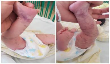
Figure 1: Multiple erythematoviolaceous macules with a reticulated pattern and well-defined, irregular borders, located on both lower limbs..
Supplementary tests
|
Parameter |
Results |
|
Complete blood count |
|
|
Hematocrit |
0,47 g/dl |
|
Platelet count |
230 x 10⁹/L |
|
Leukogram |
8 x 10⁹/L |
|
Leukocyte differential |
|
|
Neutrophils |
60 % |
|
Lymphocytes |
30 % |
|
Eosinophils |
1 % |
|
Monocytes |
3 % |
|
Basophils |
0,5 % |
|
Liver enzymes |
|
|
TGP |
31 U/L |
|
TGO |
30 U/L |
Source: Prepared by the authors.
Echocardiogram: unremarkable.
Fundoscopy: normal.
Chest X-ray: negative.
General newborn skin care measures were recommended. Additionally, an interdisciplinary evaluation was conducted, involving Cardiology, Neurology, Neonatology, and Ophthalmology, to rule out associated conditions; no organ or system damage was observed.
Based on the clinical findings (congenital reticulated lesions that blanch upon pressure, do not improve with heat, show asymmetric distribution, lack venectasias, and present no obvious extracutaneous anomalies), a diagnosis of non-syndromic cutis marmorata telangiectatica congenita (CMTC) was established. The major criteria defined by Kienast and Hoeger were met: congenital reticulated erythema, absence of venectasias, and lack of response to local warming. Minor criteria (telangiectasias, atrophy, ulceration, port-wine stain) were absent.
ICD-11 Code: 2E83.0Z (Congenital capillary malformation, unspecified – within vascular anomalies).
Treatment and management
No specific pharmacological treatment was indicated. General newborn skin care measures were established: avoiding trauma, maintaining adequate hydration with mild emollients, protecting against extreme temperatures (cold) that could accentuate the mottled pattern, and monitoring for potential ulceration or changes in the lesions.
Course and follow-up
The infant was discharged at one month of age. At 3 months of age, she attended the Provincial Genodermatosis Clinic. Dermatological physical examination confirmed the complete disappearance of the reticulated lesions, leaving no scarring, residual pigmentation, or cutaneous atrophy (Figure 2). Psychomotor development was normal for her age.
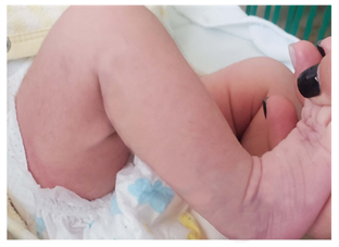
Figure 2. Complete disappearance of reticulated lesions at 3 months of age, without scarring or cutaneous atrophy.
DISCUSSION
Cutis marmorata telangiectatica congenita (CMTC) is an infrequent capillary vascular malformation, whose diagnosis is fundamentally clinical. The presented case illustrates the typical characteristics of this entity: congenital violaceous reticular lesions, which blanch with digital pressure, do not improve with local warming, present asymmetric distribution, and absence of venectasias, thus fulfilling the major criteria of Kienast and Hoeger (1,9). The rarity of this anomaly and its good course in this patient justify its communication, especially due to the complete resolution of the lesions at three months of age, which constitutes an earlier outcome than that classically described (disappearance around two years) (1,10).
In the reviewed literature, the prevalence of CMTC shows a slight female predominance, although without statistically significant differences in the published series (12). Our case, a female patient, coincides with this trend. The location of the lesions on the lower limbs, as observed in this newborn, is the most frequent, reported in up to 66 % of cases, and may also affect the trunk, upper limbs, and exceptionally the facial region (8). The asymmetric distribution, with greater involvement of the right lower limb, is another characteristic finding that supports the diagnosis (13).
The differential diagnosis must be precise to avoid confusion with entities of worse prognosis. Physiological cutis marmorata, which disappears with heat and has a symmetric distribution, was excluded in our patient upon verifying the lack of response to local warming and the persistence of the lesions in a warm environment (14). Other conditions to consider include congenital livedo reticularis, Sneddon syndrome, Klippel-Trenaunay syndrome, Sturge-Weber syndrome, and genuine diffuse phlebectasia, all of them associated with more complex vascular anomalies and often with systemic involvement (13,15). The absence of body asymmetry, bone malformations, soft tissue hypertrophy, or neurological alterations in our patient allowed these syndromes to be ruled out.
A crucial aspect in the management of CMTC is the screening for extracutaneous anomalies, which can affect up to 50–80 % of patients according to the series (5,10). The most frequently associated manifestations include body asymmetry (mainly of the limbs), delayed psychomotor development, glaucoma, congenital heart disease, cleft palate, aplasia cutis, skin atrophy, and ulcerations (10,16). In our case, a complete multidisciplinary evaluation was performed (cardiology, ophthalmology, neurology, neonatology) and complementary studies (echocardiogram, fundoscopy, chest X-ray), all negative. This approach is essential, since the early detection of associated alterations allows timely treatments to be instituted and improves the prognosis (5,14).
The course of CMTC is usually favorable, with a tendency toward attenuation or spontaneous disappearance of the skin lesions in the first years of life. Most authors report that complete resolution occurs around two years of age (1,10). However, in our patient, the total disappearance of the lesions was confirmed at three months, which constitutes an infrequent finding and of good prognosis. This data coincides with what was reported by Moreno-Alfonso and collaborators, who observed early resolution in two cases (1), and contrasts with series where the lesions persist beyond one year of life (8). No complications such as ulceration, atrophy, or bleeding were identified in our patient, which are described in a minority of cases (5).
It is relevant to compare our case with other published experiences. In a study conducted in Pamplona, Colombia, two cases with favorable course were reported, although without specifying the age of resolution (1). In contrast, Maluf Esselin and collaborators, in Brazil, found alterations in the echocardiogram and fundoscopy in a series of newborns with CMTC, which underscores the need for systematic studies (17). In our case, said examinations were normal, which reinforces the phenotypic variability of this entity.
Strengths and limitations of the case
The main strength of this report is the complete clinical documentation, with follow-up at three months and multidisciplinary evaluation that ruled out extracutaneous anomalies, as well as the photographic evidence of the lesions and their resolution. Among the limitations, we highlight: 1) the absence of genetic studies (detection of somatic mutations in GNA11), which although not necessary for diagnosis, could confirm the postzygotic somatic etiology (6); 2) the relatively short follow-up (three months), so the absence of late manifestations such as body asymmetry or later-onset glaucoma cannot be ensured; and 3) the lack of skin biopsy, which although not routinely indicated, could have been considered in the presence of atypical lesions, but was not necessary in this case.
Lessons learned
This case underscores that CMTC should be recognized early by the pediatrician, dermatologist, and neonatologist, since its accurate diagnosis avoids unnecessary interventions and allows adequate management. Although the course is usually benign, the evaluation of possible associated anomalies should not be omitted, as they can have serious consequences if not detected and treated in time. Early resolution of the skin lesions, as occurred in our patient, does not exclude the need for long-term follow-up to confirm the absence of neurological, ophthalmological, or developmental sequelae. It is recommended that every newborn with CMTC be evaluated by a multidisciplinary team and that follow-up be carried out at least during the first year of life, with periodic controls of visual acuity, intraocular pressure, and psychomotor development (5,10,14).
CONCLUSIONS
Cutis marmorata telangiectatica congenita is an entity with a good prognosis that requires early diagnosis and multidisciplinary follow-up to rule out systemic involvement. Complete resolution of the lesions may occur in the first months of life, as evidenced in this case, with total disappearance at three months of age. It is essential to differentiate it from other vascular malformations of worse prognosis and to maintain long-term follow-up to rule out late manifestations.
BIBLIOGRAPHIC REFERENCES
1. Moreno-Alfonso JC, Molina Caballero Z, Pérez Martínez E. Asociaciones infrecuentes de la cutis marmorata telangiectásica congénita: reporte de dos casos. Cir Pediatr. 2024;37:33-36. doi: 10.54847/cp.2024.01.12
2. Portal Buenaga M, Naharro Fernández C, Gómez Dermit V, De las Cuevas Terán MI. Recién nacido con cutis marmorata telangiectásica congénita. An Pediatr (Barc). 2022;97(6):433-434. doi: 10.1016/j.anpedi.2022.02.001
3. International Society for the Study of Vascular Anomalies. ISSVA Classification of Vascular Anomalies. [Internet] 2025 [cited 11/07/2026]. Available in: https://www.issva.org/classification
4. Bui TNPT, Corap A, Bygum A. Cutis marmorata telangiectatica congenita: a literature review. Orphanet J Rare Dis. 2019;14(1):283. doi: 10.1186/s13023-019-1229-8
5. Downey C, Metry D, Garzon MC, Morales LK, Baselga E. Cutis marmorata telangiectatica congenita: Incidence of extracutaneous manifestations and a proposed clinical definition. Pediatr Dermatol. 2023;40(5):820-828. doi: 10.1111/pde.15377
6. Schuart C, Bassi A, Kapp F, Wieland I, Pagliazzi A, Losch H, et al. Cutis marmorata telangiectatica congenita being caused by postzygotic GNA11 mutations. Eur J Med Genet. 2022;65(5):104472. doi: 10.1016/j.ejmg.2022.104472
7. Ghotbabadi SH, Barzegar H, Abdolvand M. Generalized cutis marmorata telangiectatica congenita or neonatal lupus? A case report and literature review. Clin Case Rep. 2024;12(5):e8898. doi: 10.1002/ccr3.8898
8. Kamińska M, Szular K, Schreiber-Zamora J, Gutowska-Malina K, Stanojević M, Kociszewska-Najman B. Newborn with cutis marmorata telangiectatica congenita: a case report and review of the literature. Pediatr Pol. 2025;100(3):276-280. doi: 10.5114/polp.2025.154298
9. Kienast AK, Hoeger PH. Cutis marmorata telangiectatica congenita: a prospective study of 27 cases and review of the literature with proposal of diagnostic criteria. Clin Exp Dermatol. 2009;34(3):319-323. doi: 10.1111/j.1365-2230.2008.03074.x
10. Boia M, Popescu DE, Jura AMC, Belengeanu V, Lungu N, Manea AM, et al. Cutis Marmorata Telangiectatica Congenita: Case Series and Literature Review. Diagnostics (Basel). 2025;15(16):2043. doi: 10.3390/diagnostics15162043
11. Manna S. A Rare Case of Vascular Malformation in India: Cutis Marmorata Telangiectatica Congenita. Cureus. 2024;16(7):e64408. doi: 10.7759/cureus.64408
12. Yorulmaz A, Hayran Y, Aktas A. Cutis marmorata telangiectatica congenita. Our Dermatol Online. 2021;12(e):e35. doi: 10.7241/ourd.2021e.35
13. Elitt MS, Tamburro JE, Moran RT, Traboulsi E. Cutis marmorata telangiectatica congenita: a focus on its diagnosis, ophthalmic anomalies, and possible etiologic factors. Ophthalmic Genet. 2020;41(2):101-107. doi: 10.1080/13816810.2020.1744018
14. Hokazono K, Urzedo ABDL, Dias PB, Dias NAL. Retinal abnormalities in a patient with cutis marmorata telangiectatica congenita. BMJ Case Rep. 2024;17(5):e257810. doi: 10.1136/bcr-2023-257810
15. Tamburro J, Traboulsi E, Patel MS. Isolated and Classic Cutis Marmorata Telangiectatica Congenita. In: GeneReviews [Internet]. Seattle: University of Washington; 2022 [cited 11/07/2026]. Available in: https://www.ncbi.nlm.nih.gov/books/NBK581081/
16. Wollina U, França K, Lotti T, Tchernev G. Van Lohuizen Syndrome – A Case Report with a Diagnostic Delay of Four Years. Open Access Maced J Med Sci. 2018;6(1):74-75. doi: 10.3889/oamjms.2018.024
17. Maluf Esselin M, Prazeres da Silva A. Cutis Marmorata Telangiectásica Congénita em RN do sexo feminino. Residência Pediátrica. [Internet] 2017 [cited 11/07/2026];7(1):36-38. Available in: https://residenciapediatrica.com.br/detalhes/255/cutis-marmorata-telangiectasica-congenita-em-rn-do-sexo-feminino
AUTHOR CONTRIBUTIONS
DBS: Conceptualization, data curation, investigation, methodology, project administration, resources, software, supervision, validation, visualization, drafting of the manuscript, writing, review, and editing of the final work.
LST: Conceptualization, investigation, drafting of the manuscript, review, and editing.
DDB: Conceptualization, investigation, drafting of the manuscript, review, and editing.
DDB: Conceptualization, investigation, drafting of the manuscript, review, and editing.
CONFLICT OF INTERESTS
The authors declare that there is no conflict of interest.
FUNDING SOURCES
The authors declare that they received no funding sources.
USE OF ARTIFICIAL INTELLIGENCE
The authors declare that generative artificial intelligence was not used for the writing, data analysis, or interpretation of results in this manuscript.